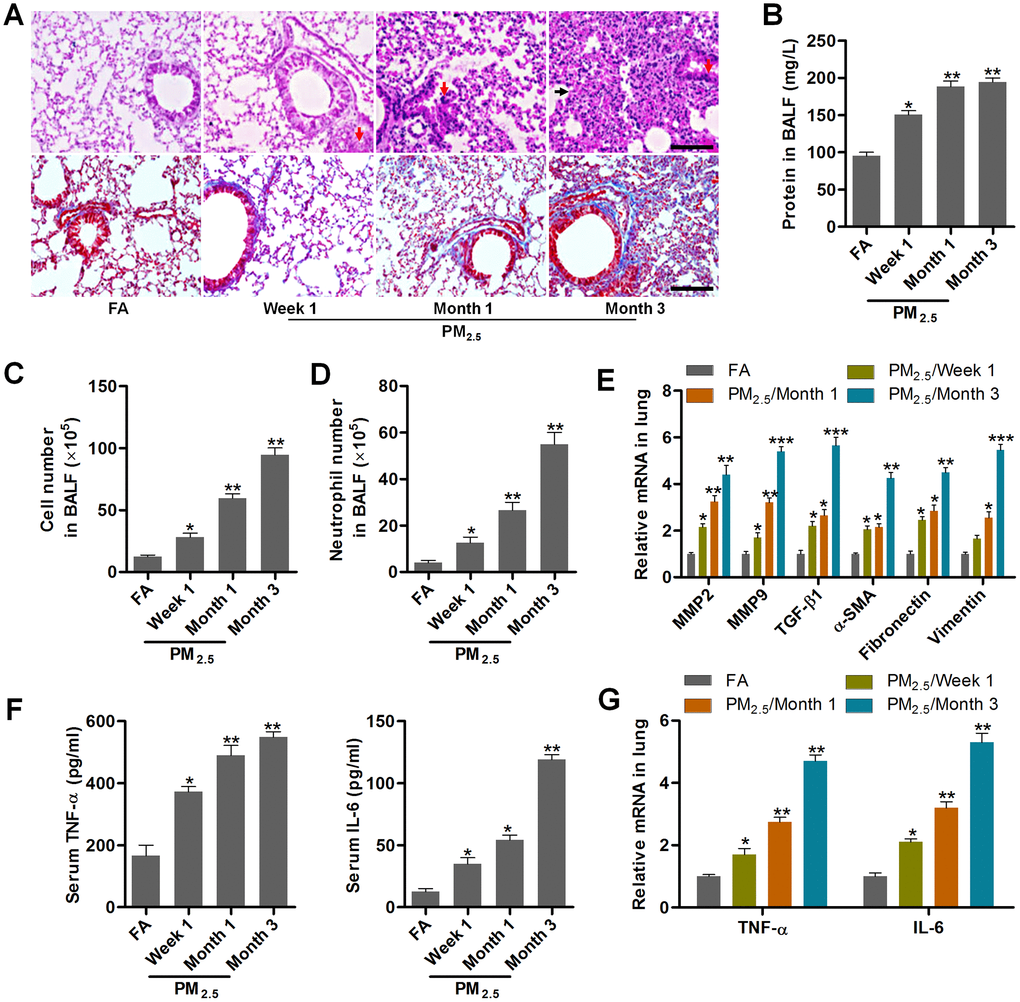
Figure 1.PM2.5 treatments result in pulmonary injury and fibrosis. (A) H&E staining (up panel) and Masson trichrome staining (down panel) of lung sections from mice challenged with PM2.5 for the indicated time points (n = 6). Scale bar, 100 μm. (B) Protein contents in the BALF were measured in mice treated with PM2.5 at the indicated time (n = 8). The number of (C) total cells and (D) neutrophils of BALF were calculated in PM2.5-challenged mice at the indicated time (n = 8). (E) RT-qPCR analysis of genes associated with fibrosis, including MMP2, MMP9, TGF-β1, α-SMA, Fibronectin and Vimentin, in lung samples of PM2.5-treated mice (n = 4). (F) TNF-α and IL-6 levels in serum of PM2.5-challenged mice were determined by ELISA (n = 8). (G) RT-qPCR analysis was used to assess TNF-α and IL-6 mRNA expression levels in pulmonary samples of mice treated with PM2.5 for the indicated time (n = 4). All data are expressed as mean ± SEM. *p<0.05, **p<0.01 and ***p<0.001 compared to the FA group.