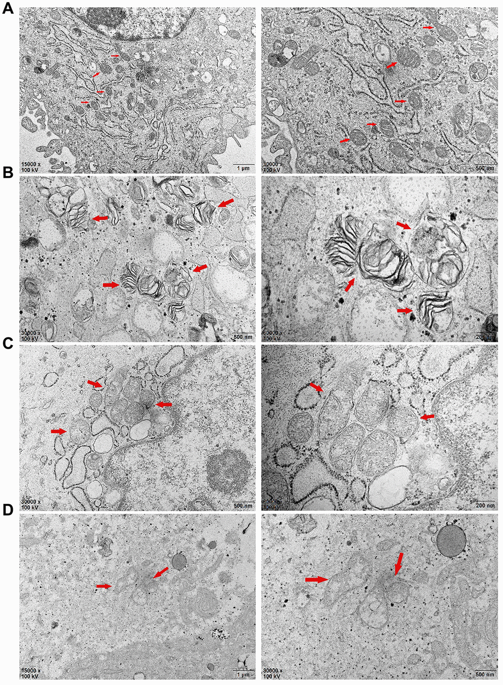
Figure 6.After 72 hours of PDLSCs culture in four different media, the microstructure changes of each group were observed by transmission electron microscopy: (A) Control Group; (B) Cells were treated with 10ng/mL TNF-α; (C) Cells were treated with 100μg/mL AGEs-BSA; (D) Cells were treated with 100μg/mL AGEs-BSA and 10ng/mL TNF-α.