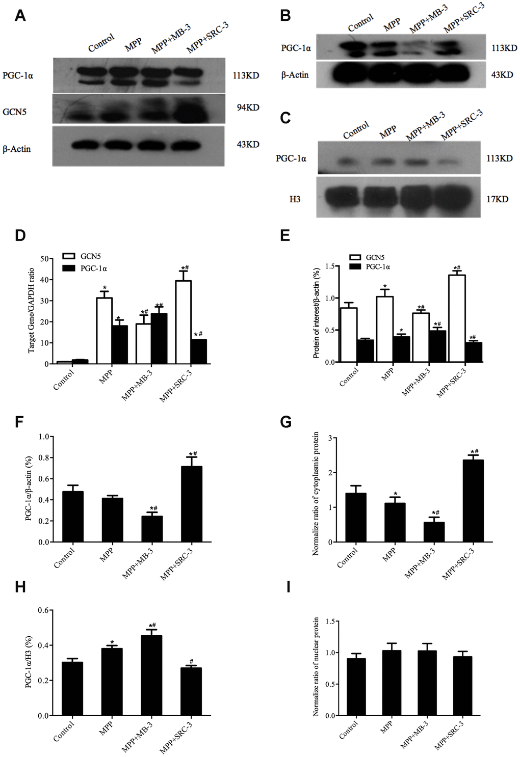
Figure 2.The cytosolic rather than the nuclear distribution of PGC-1α regulated by GCN5 in an MPP+-treated cell model. (A) The protein levels of GCN5 and PGC-1α; (B, C) The cytosolic levels of PGC-1α (B) and the nuclear levels of PGC-1α (C); (D) The relative transcriptional levels of GCN5 and PGC-1α normalized to GAPDH; (E) Semi-quantification of total GCN5 and PGC-1α proteins relative to β-actin; (F, H) Semi-quantification of the cytosolic (F) and the nuclear (H) PGC-1α proteins relative to β-actin; (G, I) The normalized cytosolic (G) and nuclear (I) proteins relative to the total protein; n=6, per group. * P <0.05, vs. Control; # P <0.05, vs. MPP+.