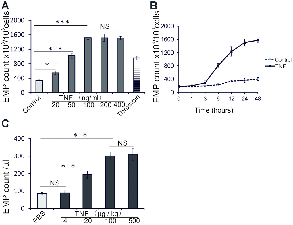
Figure 1.TNF induces EMP release in vitro and in vivo. (A) EMP release analyzed by flow cytometry (FC) in HUVECs treated 24 h with increasing TNF concentrations, or thrombin (2 IU/ml). The data represent the mean values based on 1x10⁶ cells; three independent experiments were performed. (B) EMP release analyzed by FC in untreated HUVECs, or HUVECs treated with 100 ng/ml TNF for indicated times. The data represent the mean values of EMP per 1x10⁶ cells from three independent experiments. (C) EMP release analyzed by FC in sera of TNF-treated mice. Male C57BL/6 mice (10 weeks old) were randomly assigned to 5 groups (n=10 mice per group) and treated with PBS or increasing doses of TNF for 24 hours. The data represent the mean values of EMP/μl; * P<0.05, ** P<0.01, *** P<0.001, NS indicates no significance.