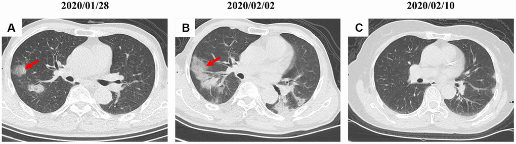
Figure 5.A serial CT images after admission of a 54-year-old male patient. (A) Patch ground-glass opacity was observed in the middle right lobe. (B) 5 days later, significant larger patch ground-glass opacities were observed in bilateral lungs. (C) Follow-up CT scans on day 13 after admission show a remarkable improvement. Typical lesions were marked with red arrows.