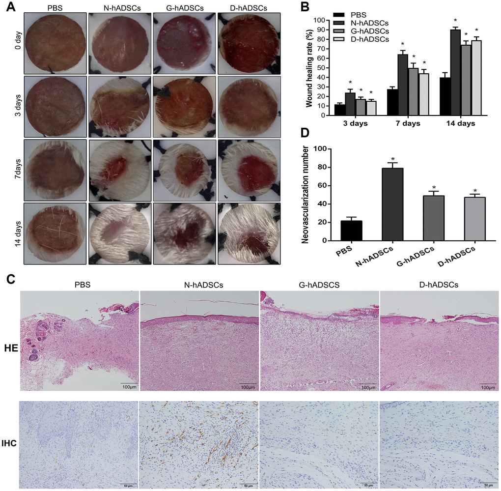
Figure 3.Glucolipotoxicity significantly reduced the treatment ability of hADSCs on skin wound healing in vivo. (A) Representative images of wound healing at time points; (B) Histogram of statistical analysis of healing rate of wounds at different time points; (C) Histology of inflammatory cell infiltration from dermis to subcutaneous layers was detected by HE staining (Scale bar = 100 μm); The content of CD31 in wound tissue was assessed by immunohistochemistry (Scale bar = 50 μm); (D) Quantification of CD31 in wound skin of different groups (* P<0.05).