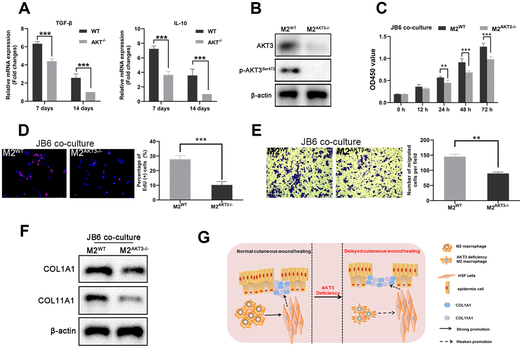
Figure 7.M2 macrophages from AKT3-/- mice failed to promote cell proliferation and migration ex vivo. (A) TGF-β and IL-10 mRNA levels were decreased in delayed cutaneous wound tissue 7th and 14th day post-injury in mice (n = 3). (B) Western blotting demonstrated the loss of AKT3 in M2 macrophages from AKT3-/- mice. (C, D) CCK-8 and EdU assays demonstrated that M2 macrophages from AKT3-/- mice were incapable of promoting JB6 cell proliferation (C) or DNA replication (D), respectively. (E) Transwell migration assay showed M2 macrophages from AKT3-/- mice could not promote JB6 cell migration. (F) COL1A1 and COL11A1 protein levels in JB6 were not increased by co-culture with M2 macrophages from AKT3-/- mice. (G) The schematic illustration of the role of M2 macrophage AKT3 deficiency in delayed cutaneous wound healing. All the experiments were repeated at least three times.