Submit an Article
Navigate
Home
Editorial Board
Editorial Policies
Current Volume
Archive
Scientific Integrity
Publication Ethics Statements
Interviews with Outstanding Authors
Newsroom
Sponsored Conferences
Podcast
Contact
Special Collections
Submit an Article
Online ISSN: 1945-4589
Research Paper
|
Volume 12, Issue 8
|
pp. 6852–6864
Swimming exercise stimulates IGF1/ PI3K/Akt and AMPK/SIRT1/PGC1α survival signaling to suppress apoptosis and inflammation in aging hippocampus
Back to article
Figure 1
(1 of 7)
−
100%
+
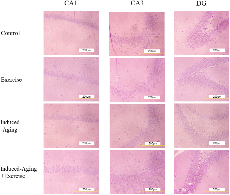
Figure 1.
Representative micrographs of H&E staining in the CA1, CA3 and dentate gyrus (DG) regions of hippocampus for each group.
The images of hippocampus architecture were magnified 400 times.