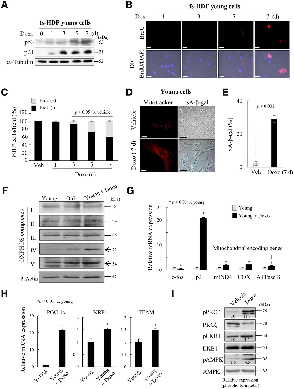
Figure 3.Mitochondria are activated in senescent fs-HDF cells. To confirm the role of PKCζ in the regulation of mitochondrial nucleoid remodeling, fs-HDF young cells were treated with doxorubicin (Doxo; 100 ng/mL) for 7 days and senescence was monitored. (A) Immunoblot analysis shows induction of p53 and p21WAF1 expression. (B) Doxo treatment induced BrdU incorporation at 5 days, the magnitude of which was greater at 7 days. BrdU-positive cells were examined in the dark, and DAPI fluorescence was observed under a differential interference contrast (DIC) fluorescence microscope (Carl Zeiss MicroImaging GmbH). Scale bars, 20 μm. (C) BrdU incorporation in mitochondria was quantified. Fluorescence microscope images were captured and counted at least 180 cells using ImageJ software (n=8 images/group). (D) Young fs-HDF cells and Doxo-treated (7 days) senescent cells were stained with MitoTracker or SA-β-galactosidase. Scale bars, 10 μm (white bar) or 50 μm (black bar). (E) Percent of SA-β-galactosidase (+) cells. Over 250 cells in 5 fields were counted. (F) fs-HDF young, old, and Doxo-treated young cells were subjected to immunoblot analysis, and then protein expressions of mitochondrial OXPHOS subunits were examined. (G) RT-qPCR analysis of the expression of complex-I, -IV, and -V subunits in young and Doxo-induced senescent cells. c-fos and p21WAF1 were used as the positive controls for proliferation and cell-cycle arrest, respectively. (H) RT-qPCR analysis of PGC-1α, NRF1 and TFAM expression in Doxo-induced senescent cells. (I) Immunoblot analysis of the expression of PKCζ, LKB1, and AMPK in Doxo-treated young cells and control cells. Band intensity was quantified using ImageJ software and normalized to amount of each protein. Effect of Doxo treatment was presented by the relative intensity of phosphorylation based on that of the vehicle treatment.
To confirm the role of PKCζ in the regulation of mitochondrial nucleoid remodeling, fs-HDF young cells were treated with doxorubicin (Doxo; 100 ng/mL) for 7 days and senescence was monitored. (J) Doxo-treated young cells were transfected with siControl or siPKCζ for 48 h and subjected to RT-qPCR analysis. Note the effect of PKCζ knockdown on mitochondrial biogenesis. (K) Fluorescence microscopy of mitochondrial nucleoid remodeling in Doxo-induced senescent cells. BrdU incorporation (red) in enlarged mitochondria was lost in the siPKCζ-transfected cells. Scale bars, 20 μm. (L) BrdU incorporation in mitochondria was quantified. Fluorescence microscope images were captured and counted at least 200 cells using ImageJ software (n=10 images/group). Data are means ± SD of three independent experiments per group. One-way ANOVA followed by Tukey HSD post hoc test.