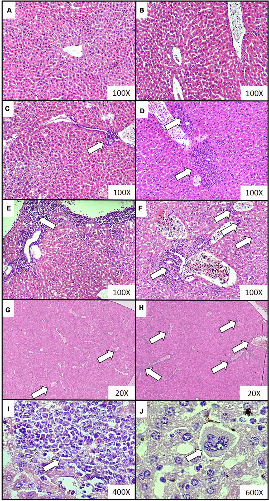
Figure 2.Representative histological images of the extramedullary hematopoiesis foci in livers of mice injected with 4T1 cancer cells. Extramedullary hematopoiesis was diagnosed histologically at increasing time intervals; observations were made based on slides from livers isolated during necropsy in animals injected with saline (control) (A) or after 0 (B), 2 (C), 3 (D), 4 (E) and 5 (F) weeks since the injection of 4T1-cancer cells. Hematoxylin and eosin staining, magnification of 100X. Additional images under lower optical magnification (20x) present extramedullary hematopoiesis foci in samples of lungs resected from mice at the second (G) and fifth (H) week of breast cancer development. The representative foci under higher magnification show the progenitor hematopoietic cells next to mature granulocytes (I) (400x) and a megakaryocyte (J) (600x). Extramedullary hematopoiesis foci are marked with white arrows. More experimental details are given in the Materials and methods section.