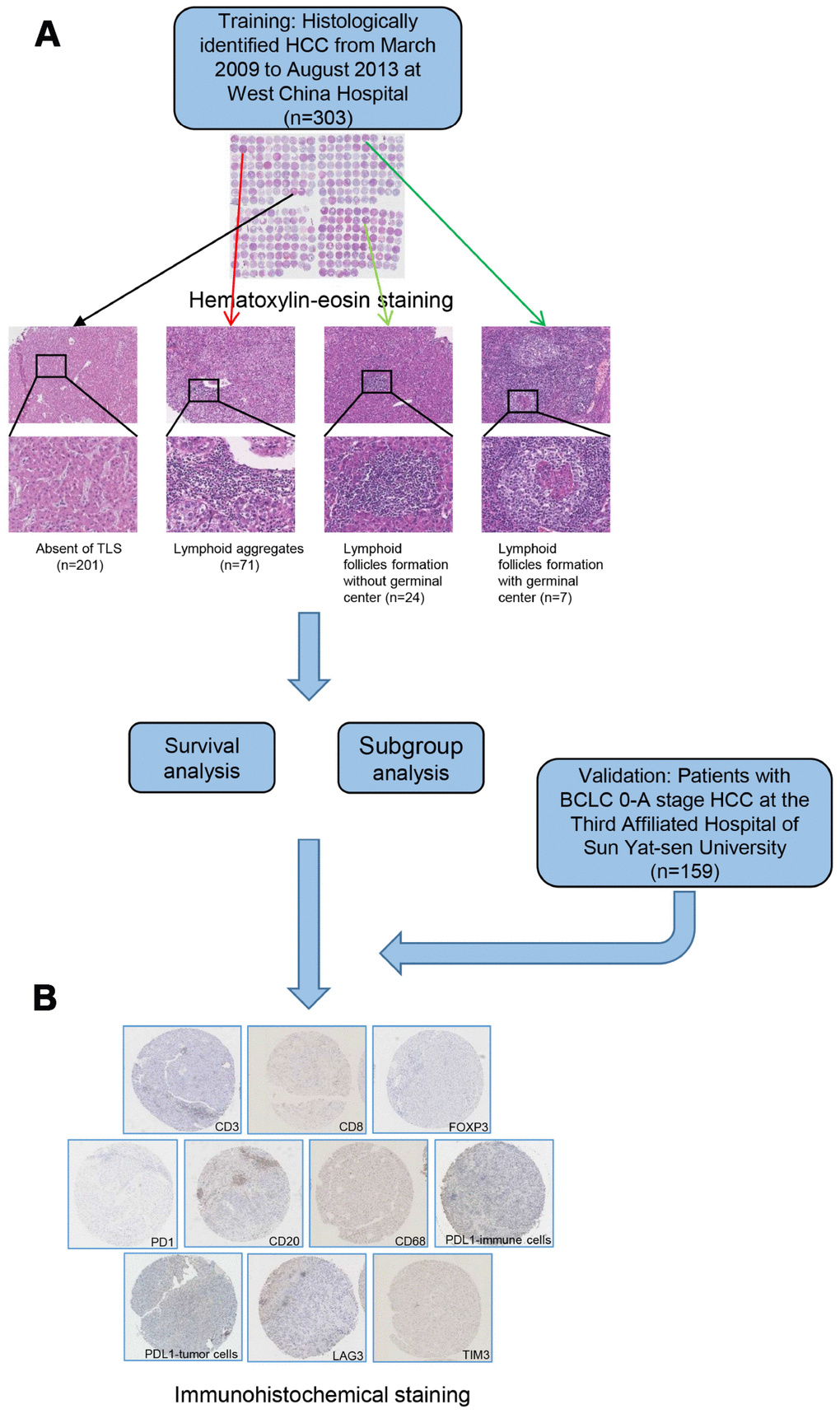
Figure 1.A flow diagram showing the characterization of intratumoral TLS. Patients were classified into 4 groups according to hematoxylin-eosin staining (A) absence of TLS; presence of lymphoid aggregates; intratumoral lymphoid follicles without germinal center; and intratumoral lymphoid follicles with germinal center. Samples from patients in training and validation cohort were examined by immunohistochemical staining (B). TLS, tertiary lymphoid structures.