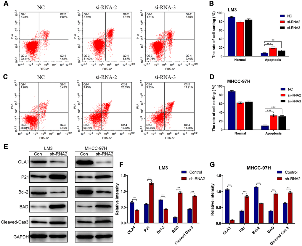
Figure 5.Depletion of OLA1 promotes apoptosis in HCC cells. (A–D) Flow cytometry was performed to determine the level of cell apoptosis in si-OLA1 groups. (E–G) Western blot analysis showed that knockdown of OLA1 upregulated P21, BAD and cleaved caspase 3 levels and downregulated Bcl-2 levels in HCC cells. All *P<0.05, **P<0.01, ***P<0.001, ****P<0.0001.