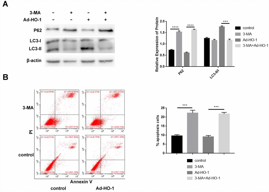
Figure 5.Autophagy inhibition by 3-MA enhances apoptosis in HO-1 overexpressing NPCs. (A) Western blot shows P62 and LC3-II/I protein levels in HO-1 overexpressing NPCs treated with or without 10 mM 3-MA. Briefly, NPCs were transfected with Ad-HO-1 for 48 h and then treated with 10 mM 3-MA to inhibit autophagy for 24 h. (B) Flow cytometry shows apoptotic rate of cells when HO-1 overexpressing NPCs were treated with or without 10 mM 3-MA. Note: All the experiments were repeated at least three times independently; ****P<0.0001 and ***p<0.001.