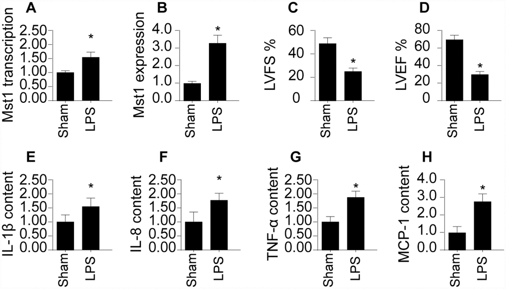
Figure 1.SRV2 is upregulated in LPS-treated cardiomyocytes. (A) RNA was isolated from LPS-treated heart tissues and qPCR was performed to analyze SRV2 transcript levels. (B) Protein was isolated from LPS-treated heart tissues, and Western blots were used to quantify SRV2 protein expression in cardiomyocytes. (C–D) Echocardiography was used to evaluate cardiac function after LPS injections. LVEF: left ventricular ejection fraction, LVFS: left ventricular fractional shortening. (E–H) Blood was collected after treatment and IL-1β, IL-8, TNF-α, and MCP-1 levels were determined using ELISA. *p<0.05 vs. control group.