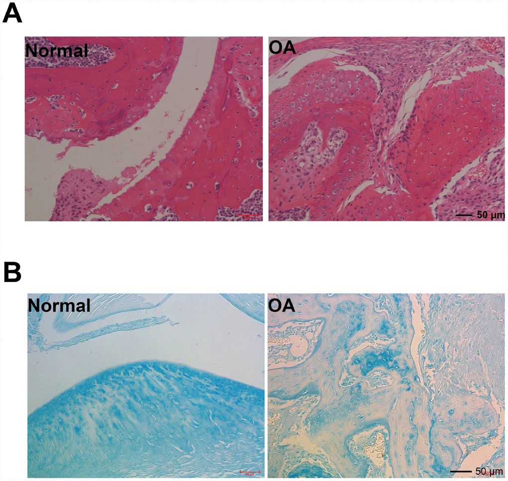
Figure 1.Hmatoxylin and eosin (H&E), and acin blue staining. (A) Representative photomicrographs HE staining of joint pathological sections of normal mouse joints and OA model mice, twelve weeks after ACLT (n=4). (B) Alcian blue staining of normal mouse joints and surgically prepared OA model mouse joint pathological sections (n=4).