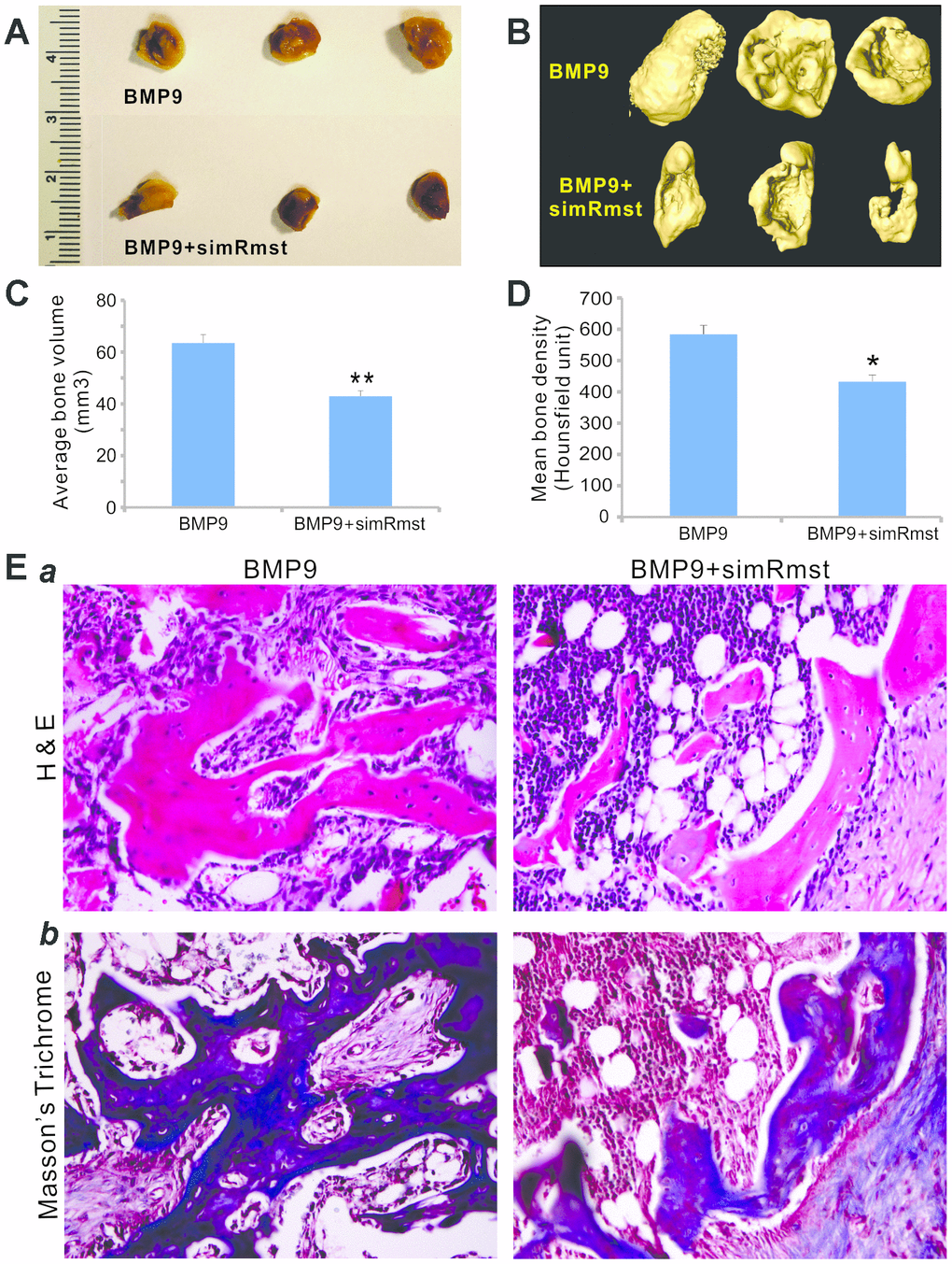
Figure 4.Silencing Rmst expression attenuates BMP9-induced ectopic bone formation. Subconfluent iMADs were infected with Ad-BMP9, Ad-GFP, and/or AdR-simRmst for 30h and collected for subcutaneous injection into the flanks of athymic nude mice. At 4 weeks after implantation, the mice were sacrificed and ectopic bone masses were retrieved. Representative macrographic images (A) and micro-CT isosurface images (B) are shown. No retrievable masses were found in the Ad-GFP or AdR-simRsmt alone group. The average bone volume (C) and mean bone density (D) were determined by analyzing micro-CT data using the Amira program. “*” p<0.05 and “**” p<0.001 Ad-BMP9 group vs. Ad-BMP9+AdR-simRmst group. (E) Histologic evaluation and trichrome staining. The retrieved masses were processed and subjected to hematoxylin and eosin staining (a) and Masson’s trichrome staining (b). Representative images are shown.