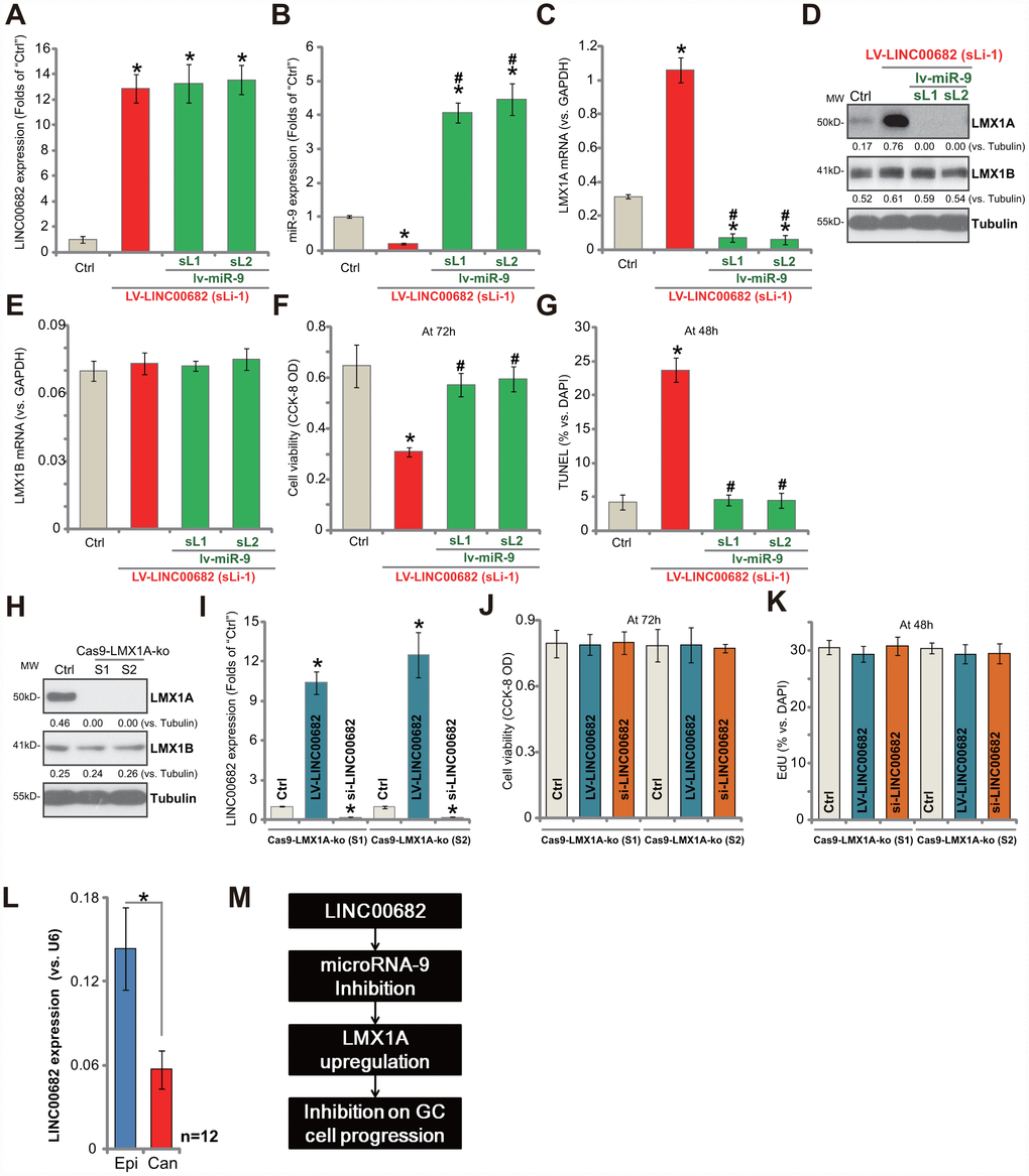
Figure 4.LINC00682 inhibits AGS cell progression via targeting miR-9-LMX1A axis. AGS cells were infected with LINC00682-expressing lentivirus (“LV-LINC00682”), following puromycin selection the stable cells were established. The stable cells (“sLi-1”) were further infected with pri-miR-9-expressing lentivirus (“lv-miR-9”) for 24h, following puromycin selection two stable lines were obtained (“sL1/ sL2”); In the cells expression of LINC00682 (A), miR-9 (B), LMX1A mRNA (C), listed proteins (D) and LMX1B mRNA (E) was tested; Cells were further cultured for applied time, and cell viability (F) and apoptosis (G) were tested by the appropriate assays. AGS cells were transfected with the lenti-CRISPR/Cas9 LMX1A knockout constructs with non-overlapping sgRNA sequences (“S1/S2”), following FACS sorting and puromycin selection two stable lines were obtained (“Cas9-LMX1A-ko”). LMX1A and LMX1B expression was tested (H). LV-LINC00682 or LINC00682 siRNA (500 nM) were transfected to the Cas9-LMX1A-ko AGS cells (“S1/S2”) for 72h, LINC00682 expression (I), cell viability (J) and proliferation (K) were tested. Expression of LINC00682 in twelve (n=12) human GC tissues (“Can”) and matched surrounding normal epithelial tissues (“Epi”) was tested by qPCR, and results were normalized to U6 RNA (L). The proposed signaling pathway of this study (M). Listed proteins were quantified and normalized to the loading control (D and H). For each assay, n=5 (five dishes or wells, except for L). *P <0.05 vs. “Ctrl” cells. #P <0.05 vs. cells without “lv-miR-9” (B, C, E–G). *P <0.05 (L). Experiments in this figure were repeated three times, and similar results were obtained.