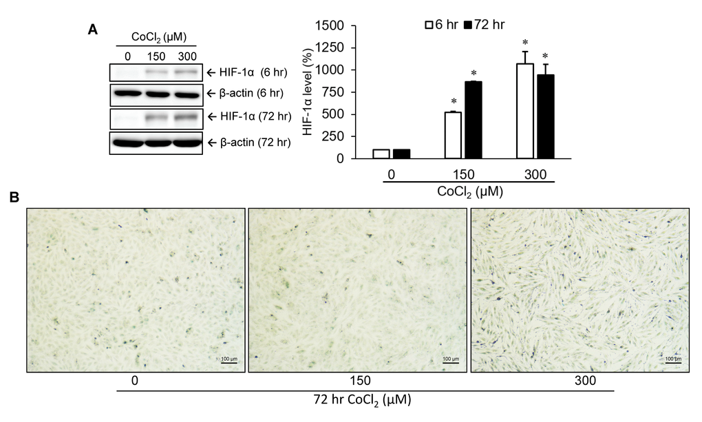
Figure 1.Identification of cellular senescence. A10 cells were incubated with various doses of cobalt dichloride (CoCl2) for 6 and 72 hr. The protein level of hypoxia-inducible factor (HIF)-1α was analyzed using Western blotting (A). Senescent cells were detected by cytochemical staining of SA-β-gal activity which appeared as a blue-green color (B). * p < 0.05 compared to the group without CoCl2 treatment.