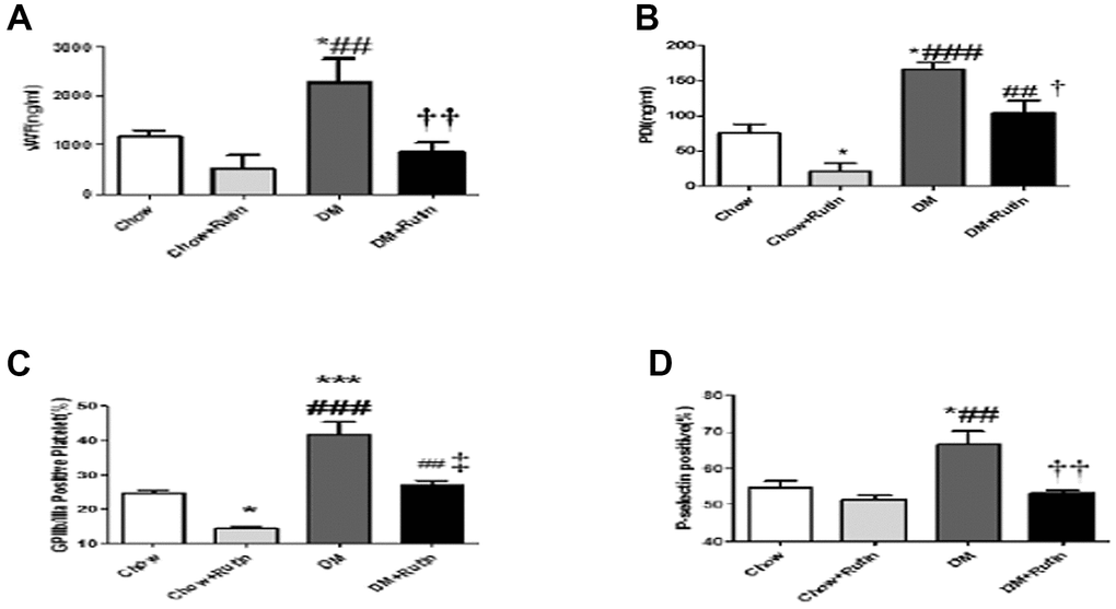
Figure 5.The plasma content of VWF and PDI was detected and the activity of platelet between the four groups was compared. (A) The VWF content in the plasma was detected using ELISA; (B) The PDI content in the plasma was detected using ELISA; (C) Flow cytometry was used to detect activated GP IIb/IIIa on the platelet surface; (D) Flow cytometry was used to detect P-selectin expression on the platelet surface *P < 0.05, **P < 0.01, ***P < 0.001, versus Chow; #P < 0.05, ##P < 0.01, ###P < 0.001, versus Chow + Rutin, †P < 0.05, ††P < 0.01, ‡P < 0.001, versus DM. Chow, normal group; Chow + Rutin, normal diet plus rutin; DM, Diabetic group; DM + Rutin, diabetic group plus rutin (n = 3–8). Data were analyzed using one-way ANOVA.