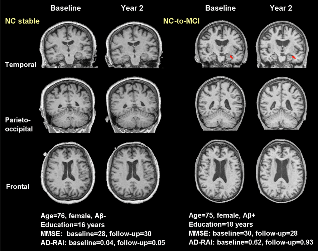
Figure 5.Typical real cases of NC stable and NC-to-MCI subjects. T1-weighted (T1W) images at baseline and two years were shown for the two typical cases in temporal, parieto-occipital and frontal view. Red arrows pointed to the region with significant atrophy by comparing the T1W images of the same subject over two years. The typical case of NC stable did not present atrophy while the case of NC-to-MCI showed increased width of left choroid fissure and temporal horn (temporal view). Aβ-: CSF-based Aβ42 >192 pg/ml at baseline and 2 years; Aβ+: CSF-based Aβ42 <192 pg/ml at baseline and 2 years; AD-RAI: AD resemblance atrophy index.