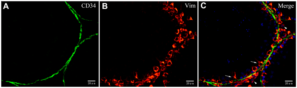
Figure 10.Double immunofluorescence staining of telocytes in rat testis with anti-CD34 and anti-vimentin antibodies. (A–C) Immunofluorscence staining shows that CD34 (green) and Vimentin (red) do not co-localize around the seminiferous tubule. Vimentin is expressed on Sertoli cells (pointed by arrows) and Leydig cells (pointed by triangles). Scale Bar = A–C: 20μm.