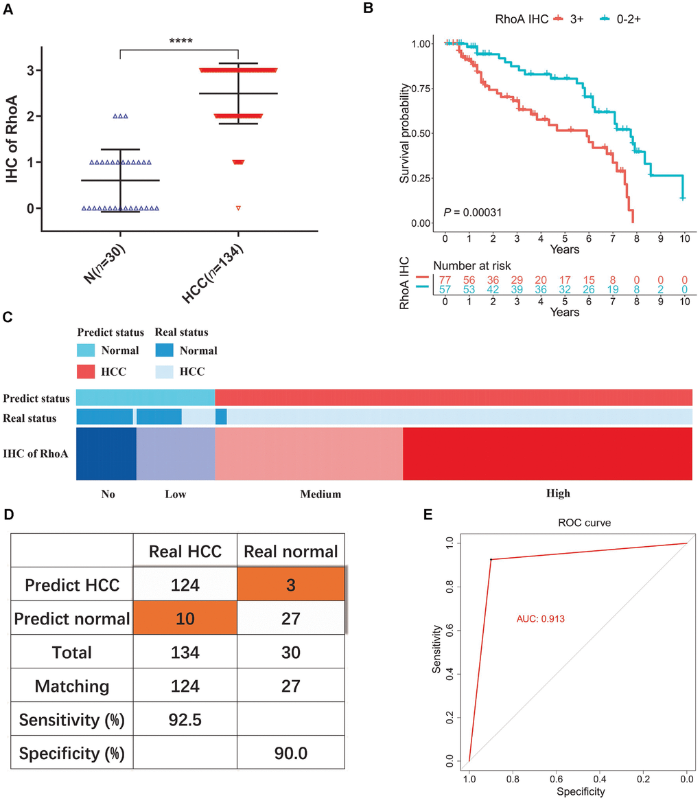
Figure 4.Diagnostic model of RhoA protein expression in liver cancer patients. The staining intensities of RhoA via immunohistochemistry chips from PUMCH patient samples (A). Kaplan-Meier curves of overall survival (B) of liver cancer patients with high RhoA protein expression levels (3+) and low RhoA protein expression levels (0–2+). Diagram (C), sensitivity and specificity validation (D) and receiver operating characteristic curve (E) of the diagnostic model according to RhoA immunohistochemistry level.