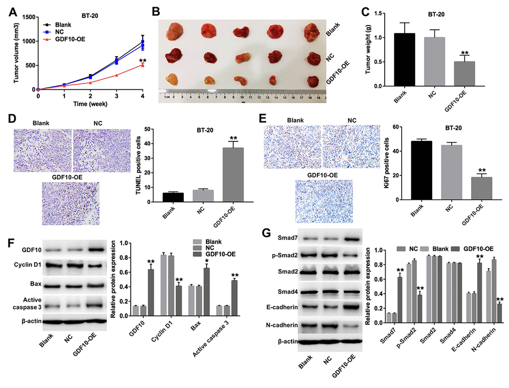
Figure 6.GDF10 expression inhibits BT-20 xenograft growth. (A) Tumor volumes were measured weekly post-inoculation of BT-20 cells infected with lentiviruses carrying the GDF10 gene or non-coding controls (NC). **P < 0.01, compared with the NC group. (B) Photographs of BT-20 xenografts dissected 4 weeks after tumor cell inoculation. (C) Tumor weights. **P < 0.01, compared with the NC group. (D) TUNEL staining of BT-20 tumors and quantification of TUNEL-positive cells. **P < 0.01, compared with the NC group. (E) Ki67 IHC in excised BT-20 tumor sections and quantification of Ki67-positive cells. **P < 0.01, compared with the NC group. (F) GDF10, cyclin D1, Bax, and active caspase-3 expression was assessed in tumor samples by western blotting. β-actin was used as internal control. Relative protein expression levels were quantified by densitometry and normalized to β-actin. *P < 0.05, **P < 0.01, compared with the NC group. (G) The expression of Smad7, p-Smad2, Smad2, Smad4, E-cadherin, and N-cadherin was investigated by western blotting in excised tumor samples. β-actin was used as internal control. Relative protein expression levels were quantified by densitometry and normalized to β-actin. *P < 0.05, **P < 0.01, compared with the NC group.