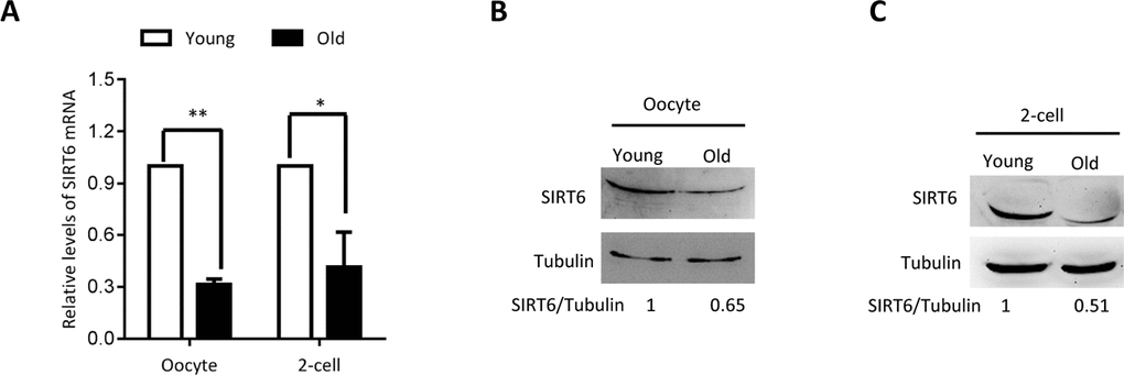
Figure 1.SIRT6 expression in aged mouse oocytes and embryos. Fully-grown oocytes and two-cell embryos from young and aged mouse were collected to evaluate SIRT6 expression. (A) Quantitative RT-PCR showing the lowered SIRT6 mRNA levels in aged oocytes and two-cell embryos. (B–C) SIRT6 protein expression in aged oocytes and two-cell embryos was verified by immunoblotting. Tubulin served as a loading control. Band intensity was calculated using ImageJ software, and the ratio of SIRT6/tubulin expression was normalized and values are indicated. Data are expressed as mean percentage ± SD of three independent experiments. *P <0.05, **P <0.01.