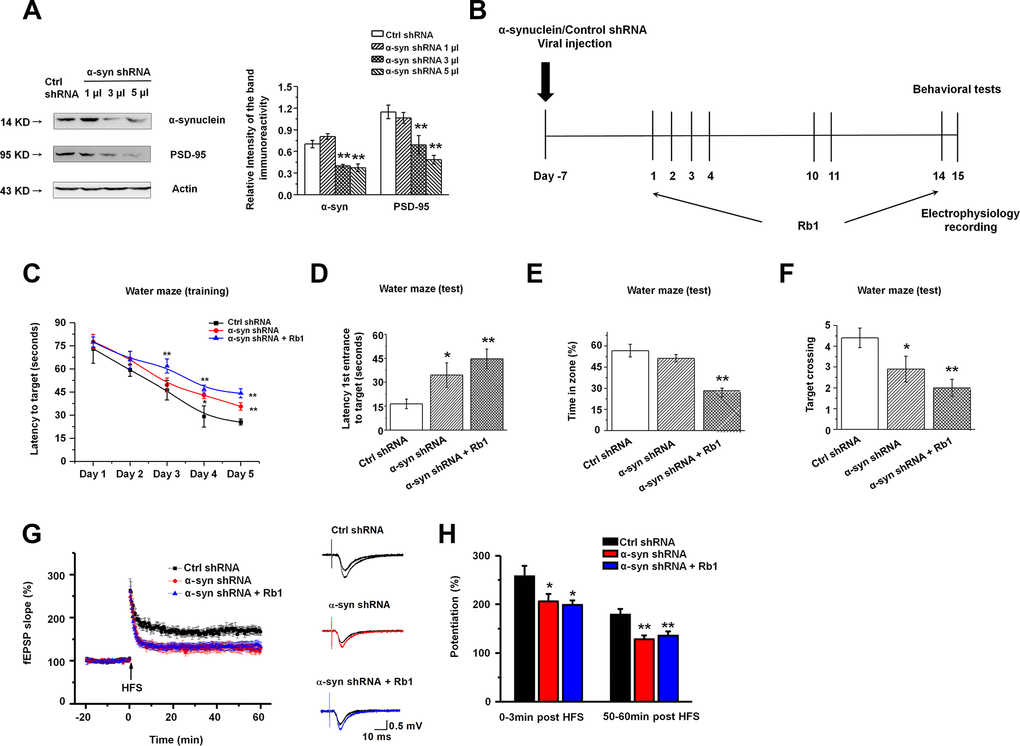
Figure 5.Knockdown of α-synuclein in hippocampal CA3 impaired learning and memory in normal mice. (A) The interference efficiency of α-synuclein shRNA was confirmed by Western blotting. n = 6 per group. (B) Experimental timeline. Seven days after the LV- α-synuclein shRNA or control shRNA virus was stereotaxically injected in the hippocampal CA3 region, mice were given saline or Rb1. Control shRNA and α-synuclein shRNA mice were intraperitoneally injected with vehicle (saline) from day 1 to day 14. α-Synuclein shRNA+Rb1 mice were intraperitoneally injected with Rb1 from day 1 to day 14. One day after the last Rb1/saline injection (day 15), behavioral tests and electrophysiological recording were performed. (C–F) Morris water maze tests were conducted after treatment with α-synuclein shRNA and Rb1. Mice were analyzed for (C) the escape latency during a 5-day training course. In the probe tests, mice were analyzed (D) for the escape latency, (E) the time spent in the target zone, and (F) the target crossing to reach the target platform from the entrance. n = 12 per group. (G) LTP at the SC-CA1 synapses was recorded in mice treated with α-synuclein shRNA or α-synuclein shRNA + Rb1. The middle image shows representative traces of fEPSP recordings of responses before and 50 min after high-frequency stimulation (HFS; arrow). (H) Quantitative analysis of LTP data in (G). The level of fEPSP potentiation was determined at a mean of 0–3 min and 50–60 min after high-frequency stimulation. n = 6. Results are expressed as the mean ± SEM. **p < 0.01, *p < 0.05 vs. control shRNA group. Statistical significance was determined by one-way ANOVA and Bonferroni tests as post hoc comparisons.