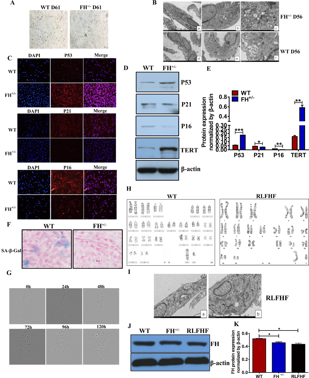
Figure 6.Further characterization of the FH+/– cells(A) Microscopic images of WT and FH+/– cells at the 61st passage (200×). (B) Representative ultrastructure images of WT and FH+/– cells at the 55th passage evaluated by electron microscopy (a,d:2000×; b,e:4000×; c.f:8000×), where a larger cell body with plenty of rough endoplasmic reticulum, mitochondria and ribosomes is shown in the FH+/– cells, but the WT fibroblasts show fewer mitochondria and ribosomes and deformed and less condensed rough endoplasmic reticulum. (C) Representative immunofluorescence images of the expression of p53, p21 and p16 (200×). Western blot for p53, p21, p16 and TERT in WT and FH+/– cells and the corresponding histograms are shown in (D) and (E), respectively. (F) SA-β-Gal detection . (G) FH+/– single cell cloning images captured with the Incucyte Zoom System for cells at the 84th passage. (H) The karyotype of a RLFHF FH+/– cell, where no chromosome abnormality is seen as revealed for the karyotype of a control fibroblast at the 40th passage. (I) Electron microscope images of RLFHF FH+/– cells (4000× for both a and b), which are rich in mitochondria, rough endoplasmic reticulum and ribosomes. Western blot for FH protein expression in the 58th passage WT, 68th passage FH+/– and RLFHF cells is shown in (J) and corresponding histograms are shown in (K).