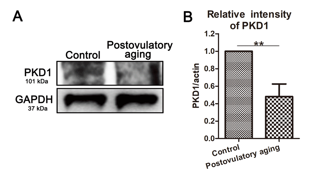
Figure 4.PKD1 expression decreases in aged oocytes. (A) Protein levels of PKD1 in control and postovulatory aging porcine oocytes were determined by western blotting. Rabbit polyclonal anti-PKD1 antibody was adopted. (B) Quantitative analysis of PKD1 expression in control and aging groups. Data are presented as mean ± s.d. from at least three independent experiments. **, significant difference (P < 0.01).