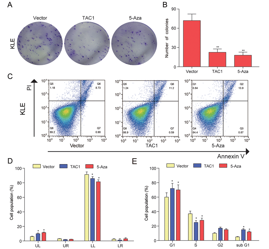
Figure 9.TAC1 demethylation inhibited KLE cells proliferation, arrested the cell cycle at the G1, and accelerated cell apoptosis. (A-B) Colony formation assay was used to assess cell proliferation ability and proliferation was indicated by number of colonies. (C-D) Cell apoptosis was determined by Annexin V/PI double staining and percentages of cells in each phase (LL, viable; LR, early apoptotic; UL and UR, late apoptotic/necrotic cell) were calculated. (E) Relative cell numbers in each cell-cycle phase (G1, S, G2, and subG1) were determined by flow cytometer of the TAC1 and vector control. *P<0.05, **P<0.01, compared with corresponding control group.