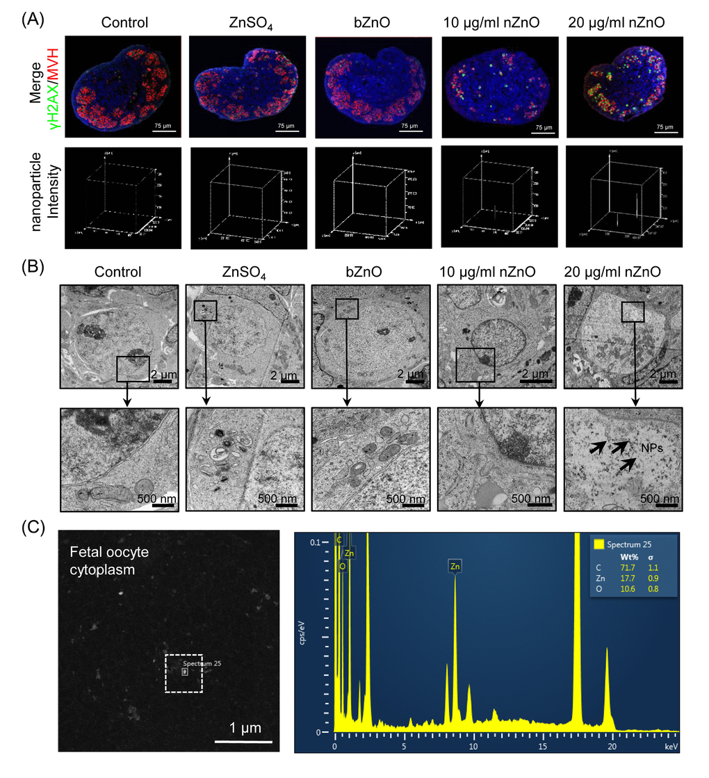
Figure 1.Internalization of nZnO in fetal oocytes. (A) Up: confocal reflection imaging of tissue sections of ovaries cultured for 6 days stained for MVH (red) and Hoechst 33342 (blue); nZnO was reflected and visualized as white dots. Down: 3-D plotting of nZnO intensity is presented with the z-axis indicating the intensity of nZnO within the ovary section and the interface of the x- and y-axes indicating the whole ovary section. (B) Ovary sections observed at TEM; NPs are not detectable in the control, ZnSO4 or bZnO whereas nZnO are recognizable as black particle in the oocyte cytoplasm (arrows). N: nucleus; C: cytoplasm. (C) Chemical characterization of nZnO nanoparticles with TEM equipped with energy dispersive spectrometer (EDX) within the oocyte cytoplasm.