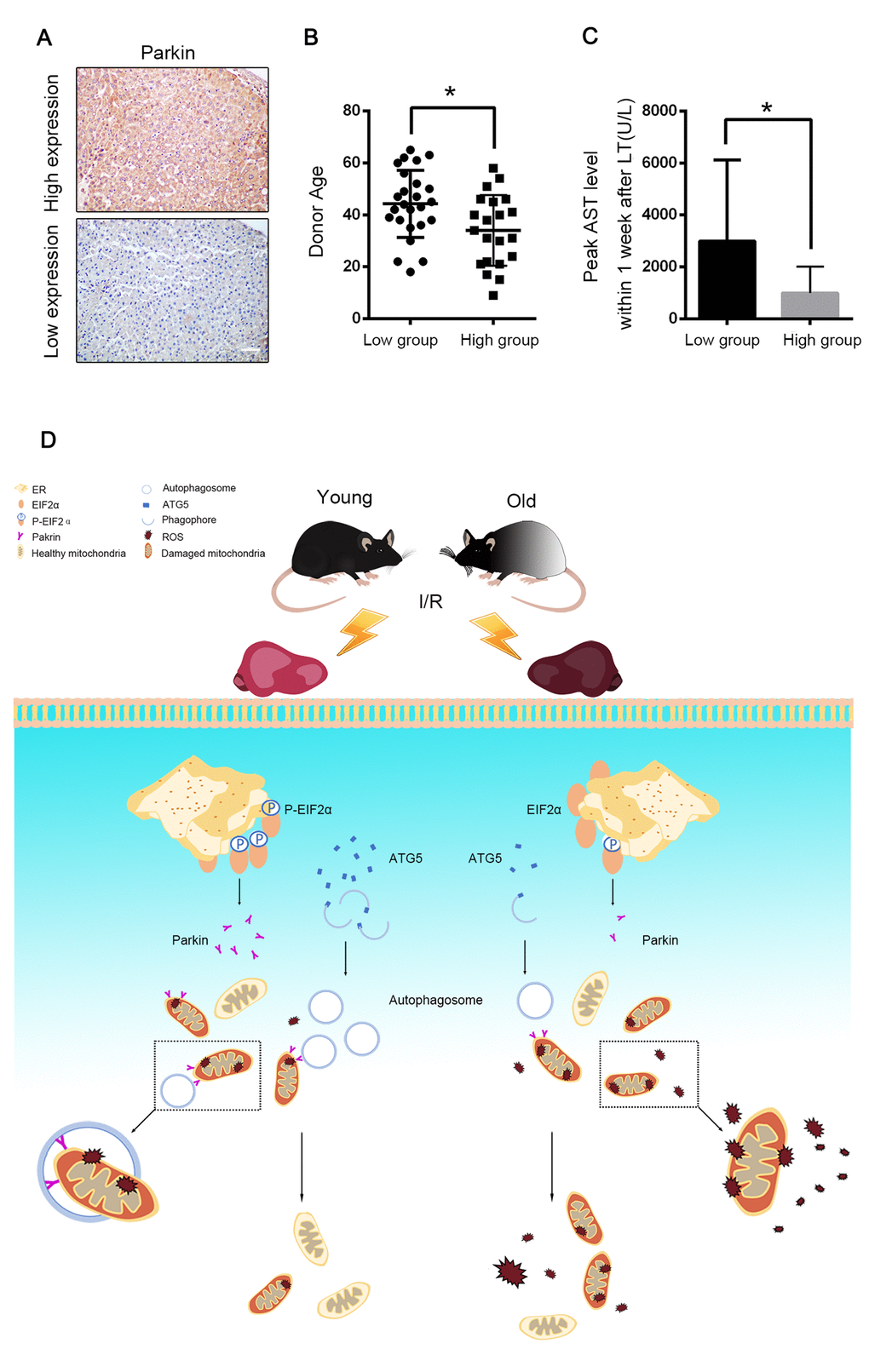
Figure 8.Parkin predicted allograft I/R injury after liver transplantation. 46 graft biopsies were performed 2 hours after complete revascularization in 46 patients undergoing DCD liver transplantation. The data of donor age and peak AST within 7 days after transplantation were collected. (A) Representative images of Parkin expression in liver graft by immunohistochemistry, 21 patients were in Parkin high-expression group and 25 patients were in Parkin-low expression group. Scale bar: 50μm. (B) The donor age of low-expression group was significantly older than high-expression group (44.2±2.6 vs. 34.0±3.0, p<0.05.) (C) The peak AST within 7 days after transplantation of low-expression group were significantly higher than high-expression group (2991±624.4 U/L vs. 993.6±221.8 U/L, p<0.01). (D) Aging aggravated hepatic I/R injury by impairing age-dependent mitophagy function via insufficient Parkin and Atg5 expression. Atg5 decreases in old reperfused liver leading to less formation of autophagosomes. Reperfusion of old ischemic mice liver decreases phosphorylation of EIF2α, which in turn inhibits Parkin expression. Reduced parkin expression and autophagosomes formation subsequently impairs mitophagy and promotes onset of the MPT and cell death. Atg5 and Parkin deficiency is responsible for age-dependent mitophagy impairment.