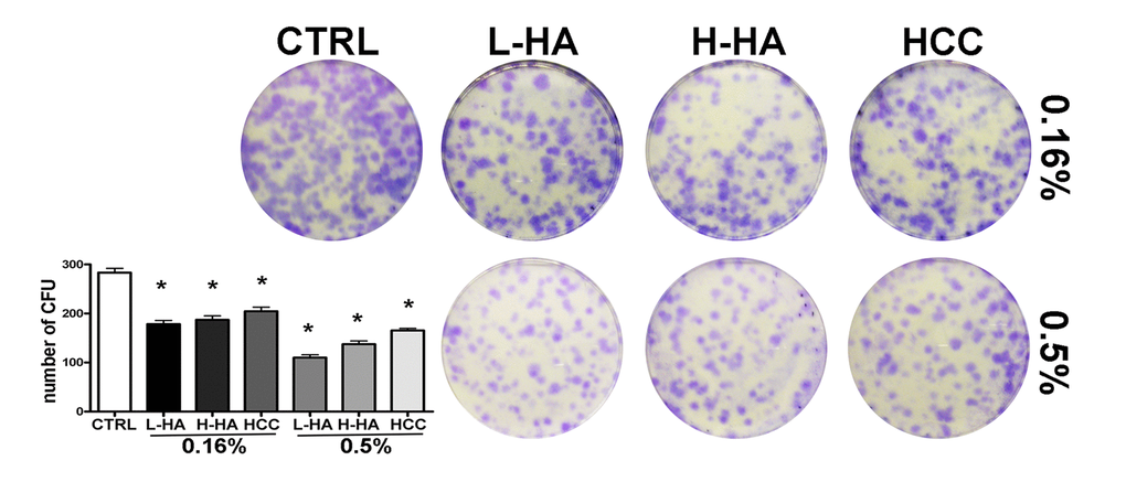
Figure 3.CFU assay. The pictures show representative crystal violet staining of clones obtained after 14 days of incubation with MSCs plated following treatment with different HA solutions. The mean number of clones (± SD, n = 3, *p<0.05) is indicated in the histogram.