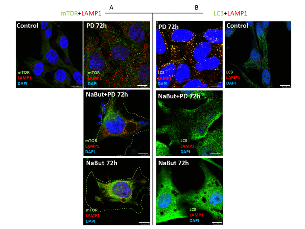
Figure 7.Spatial uncoupling of lysosomes and autophagosomes in senescent cells blocks their fusion upon activation of autophagy by MEK-ERK inhibition. Immunofluorescent images showing LAMP1 (red) and mTOR (green) perinuclear localization in senescent cells and in senescent cells upon MEK/ERK suppression. Cells were treated with inhibitors for 72 h, then fixed and stained with antibodies against LAMP1/mTOR (A) and LAMP1/LC3 (B). Green dotted lines show borders of senescent cells (A). Right panel (B) shows the actual size of senescent cells according to LC3 fluorescence. LAMP1 colocalizes with LC3 in control cells exposed to PD and does not colocalize in senescent cells. Nuclei stained with DAPI (blue). Scale bars: 7,5 µm.