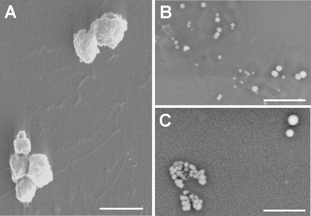
Figure 5.Scanning electron microscopy. (A) Gold-palladium coated senescent MVs. Scale bar 1μm. Note the poly-L-lysine layer. (B and C) Young MVs prepared without metal coating. Note the heterogeneous size and brightness. Scale bar 5μm. The background noise of some pictures a consequence of the fast scanning speed.