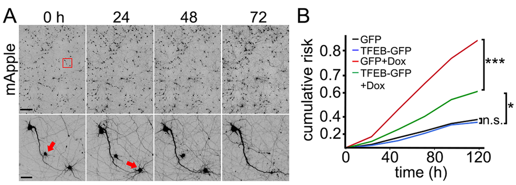
Figure 5.TFEB increases survival of doxorubicin-treated neurons. (A) An example of longitudinal imaging and survival analysis. Primary cortical neurons transfected with mApple were followed with an automated microscope. Images collected every 24 h demonstrate the ability to return to the same field of neurons and track them over time. Each image is a montage of images captured in the center of one well of a 24- or 96-well plate. Scale bar is 400 μm. In the lower panel, images are zoomed in. Arrows represent two neurons that degenerate over time. Scale bar is 50 µm. (B) Two cohorts of primary cortical neurons transfected with mApple + GFP were treated either with a vehicle or 10 nM doxorubicin. Two cohorts transfected with mApple + TFEB-GFP were treated either with a vehicle or 10 nM doxorubicin. 16 hours after treatment, the four cohorts of neurons were imaged and tracked over 5 days. Risk of death associated with doxorubicin was calculated with JMP software. *p<0.01, ***p < 0.0001 (Log-Rank test), n.s., non-significant. One hundred fifty neurons were analyzed per condition. Results were pooled from two independent experiments.