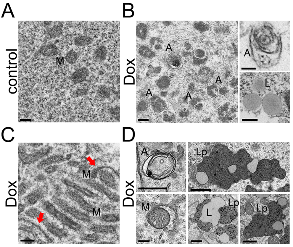
Figure 3.Accumulation of autophagosomes and organelles in primary cultured neurons and mouse brains induced by doxorubicin and doxil, respectively. (A) An example of an electron micrograph of cultured cortical neurons treated with a vehicle (overnight). M, mitochondria. Bar, 200 nm. (B) Electron micrographs of cultured cortical neurons treated with 50 nM doxorubicin (overnight). Left panel: A, autophagosomes. Bar, 200 nm. Right upper panel: A, an autophagosome. Bar, 100 nm. Right lower panel: L, lipid droplets. Bar, 500 nm. (C) An example of an electron micrograph of cultured cortical neurons treated with 50 nM doxorubicin (overnight). Note an abnormally large cluster of mitochondria. Arrows note atypical mitochondria. Bar, 200 nm. (D) Electron micrographs of mouse brain exposed to doxil. Left panels: A, autophagosomes. M, a mitochondrion being engulfed by an autophagosome. Bar, 200 nm. Right panels: L, lipid droplets; Lp, lipofuscin. Bar, 500 nm.