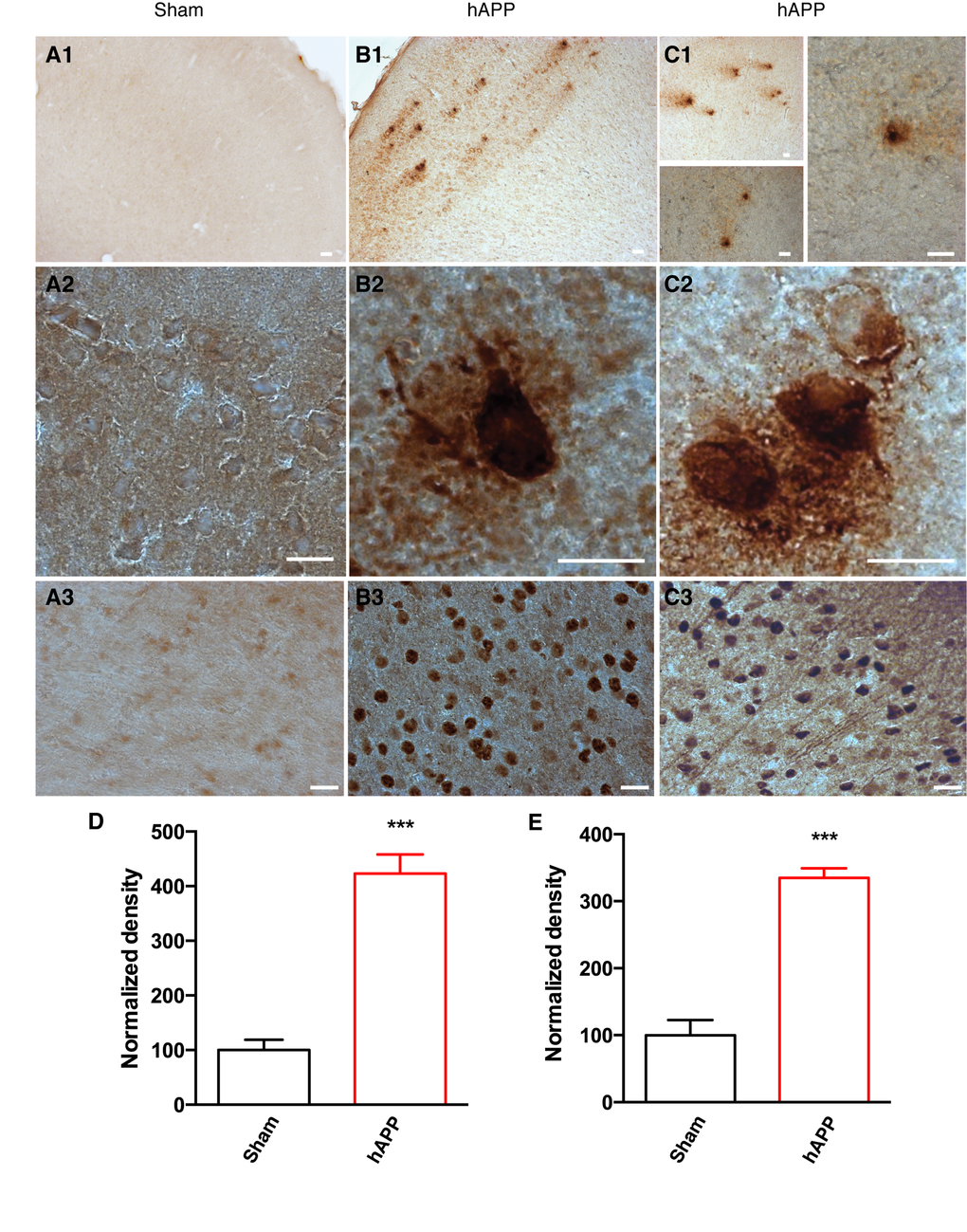
Figure 4.Detection of amyloid plaques and NFTs in the PFC of AAV-hAPP-SLA injected mice at 12 mpi. (A) Sham mouse brains injected with a control AAV and (B), (C) AAV-hAPP-SLA injected mice stained with the 4G8 antibody (1, 2) and AT100 antibody (phospho-tau at Ser212 and Thr214) (3). Scale bars = 20 µm. Quantification of 4G8 optical density (D) and AT100 densities (E) and normalized to values obtained in sham operated mice. The error bar is ± SEM. (Student’s test, P < 0.001, for the quantification of amyloid plaques, 8 slices were analyzed from 4 control mice and 6 slices from 3 AAV-hAPP injected mice, whereas for the quantification of NFTs, 6 slices were analyzed from 3 control mice and 6 slices from 3 AAV-hAPP injected mice).