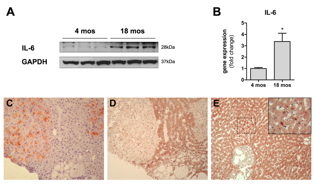
Figure 5.Panels (A) and (B) Expression of IL6 in the liver of animals transplanted with pre-neoplastic hepatocytes in at young or old age and killed 3 months after Tx. Both the protein (panel A) and the corresponding mRNA (panel B) were expressed at higher levels in the liver of aged animals. Panels (C) and (D) serial sections showing histochemical staining for DPPIV+ (panel C) and immuno-histochemical staining for STAT3 (panel D) in clusters of transplanted nodular hepatocytes. Only rare STAT3+ nuclei were detected in the aged rat liver (panel E). 100x magnification.