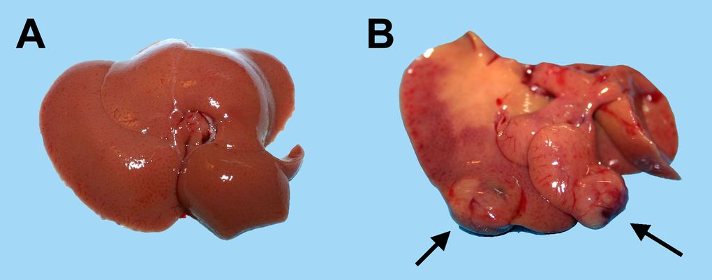
Figure 2.Macroscopic appearance of livers from animals transplanted with pre-neoplastic hepatocytes in at young or old age and killed 8 months after Tx. Panel (A) liver from a rat transplanted at 5 months of age and killed 8 months later: liver surface is regularly smooth and no lesions were detected. Panel (B) liver from a rat transplanted at 20 months of age and killed 8 months later: note the presence of two large nodules with prominent vasculature (arrows).