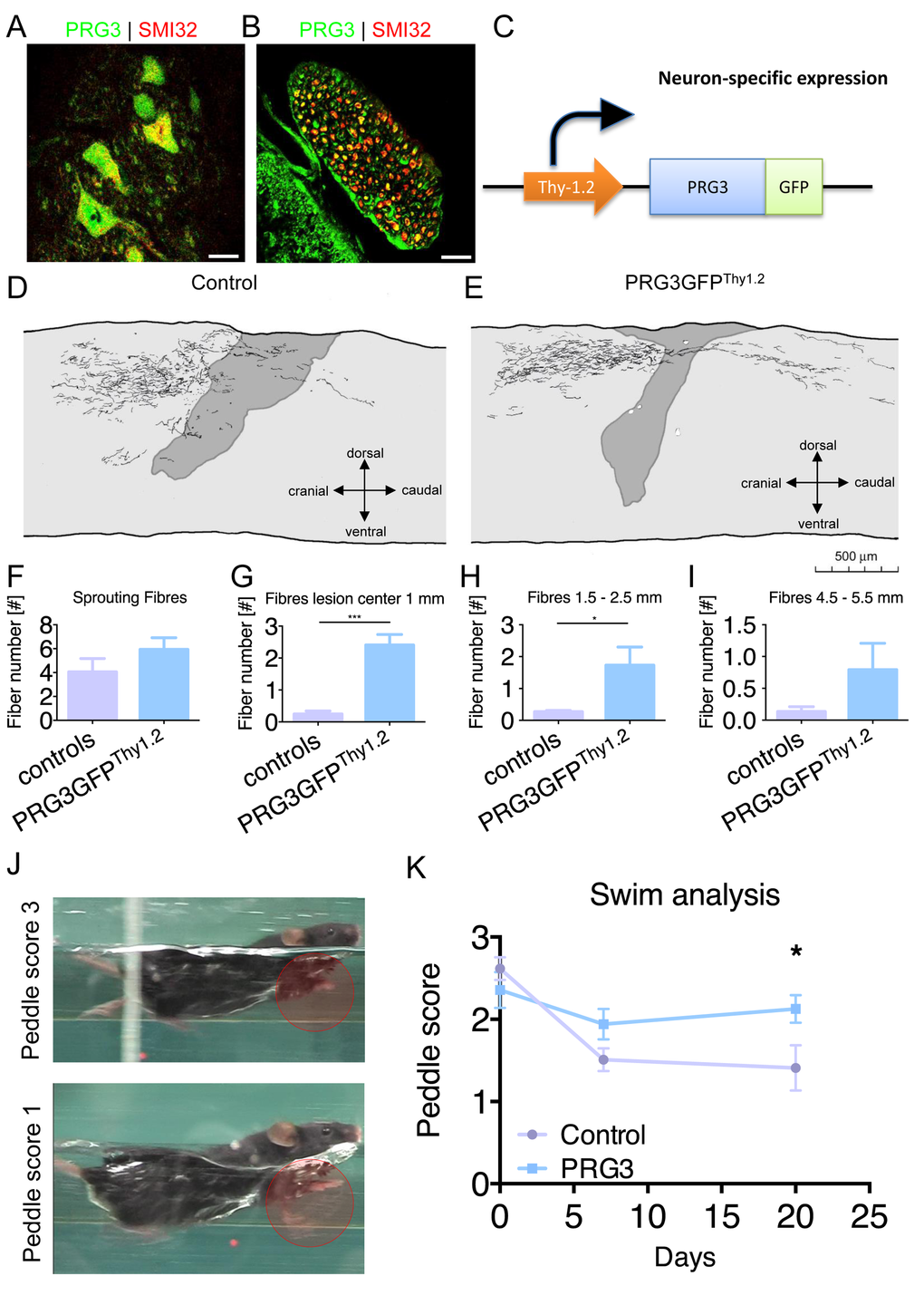
Figure 9.PRG3 is present in spinal cord neurons and contributes to spinal regeneration after spinal cord injury. (A-B) Double-immunocytochemistry showing that PRG3 is co-expressed with the motor neuron marker SMI32 (non-phosphorylated neurofilament) in murine spinal cord motor neurons in the ventral horn (A) and their axons in the ventral root (B). Scale: 20 µm. (C) Schematic illustration of the Thy1.2 driven PRG3GFP transgenic mouse, cDNA construct of PRG3-GFP was subcloned downstream of the artificial Thy1 neuronal specific promoter, the construct was introduced as transgene in 129S zygotes. (D-E) Camera lucida reconstruction of spinal cord hemisection performed in control n=4 (D) and PRG3 transgenic n=3 (E) animals. (F-I) Sprouting fibres (F) and fibres 1 mm (G), 1.5 – 2.5 mm (H) and 4.5 – 5.5 mm (I) rostral to the lesion analysis of control and PRG3 transgenic animals after dorsal hemisection based on camara lucida reconstructions. (J) Representative image of the swim test score 3 and 1 display functional locomotion recovery analysis. (K) Swim test analysis. Baseline swim analysis was performed before dorsal hemisection (time point 0). After 7 and 20 days, swim performance was again scored. PRG3 transgenic animals show significant increased swim performance 20 days post spinal cord lesion compared to controls. Data plotted as mean ± SEM. Statistical differences were analysed by two way anova with Bonferroni post hoc analysis including repeated measurement correction (n=5 per group). P value was set as * = p<0.05: ** = p<0.01; *** = p<0.001.