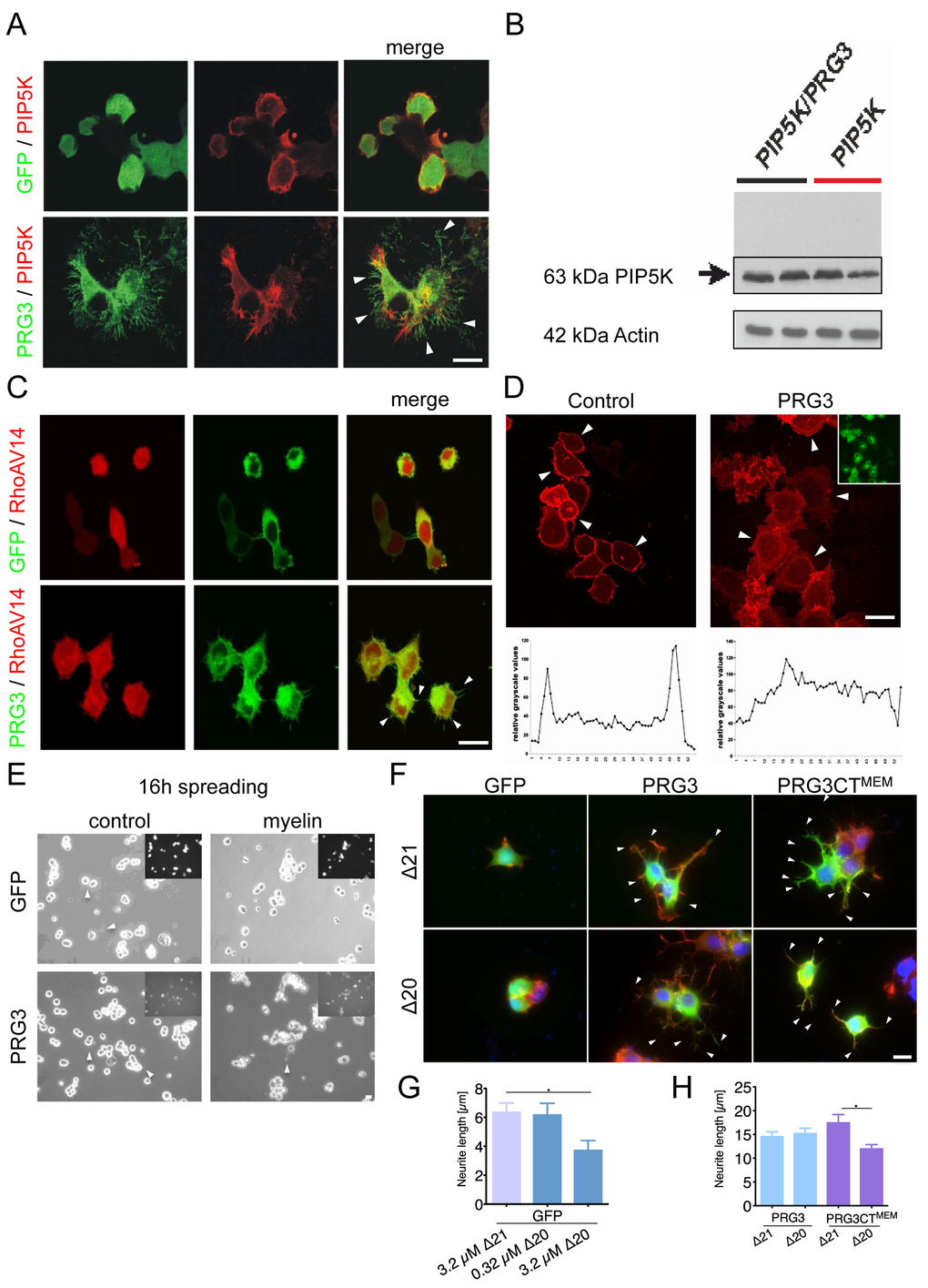
Figure 5.PRG3 impedes myelin and RhoA-induced axon collapse and translocates PIP2 from the plasma membrane. (A) Representative images of neurons expressing GFP and PIP5K (control) or PRG3 and PIP5K (PRG3). PRG3-expressing neurons still retain their complex morphology in the presence of PIP5K overexpression (arrows). Scale bar represents 20 µm. (B) Immunoblot for PIP5K expression in PRG3 expressing neurons. Bottom, Actin serves as a house keeping protein for equal gel loading. (C) Representative images of co-overexpression of RFP and PRG3 in the presence of dominant active RhoAV14. Note the increased filopodia in PRG3 expressing neurons (arrows). Scale bar represents 20 µm. (D) Disruption of phosphoinositol-(4,5)-bisphosphate (PIP2) in PRG3 expressing neurons. Representative pictures of GFP (control) and PRG3 overexpressing (PRG3) neurons expressing the RFP – PLC1 PH domain fusion constructs (red) as an in vivo probe for intracellular PIP2 localization. Transfection efficacy is given in the upper right corner. Bottom, representative trans-cellular pixel traces of PIP2 values in GFP (control) and PRG3 overexpressing (PRG3) neurons. In PRG3 cells PIP2 membrane dislocation could be observed (arrows). Scale bar represents 20 µm. (E) Axon spreading assay on myelin-coated substrates. Representative images of a spreading assay with GFP and PRG3 overexpressing neurons on control substrate and myelin substrate. Note that PRG3 expressing neurons form neurites after 16 hours on myelin. (F) Representative images of control (GFP), PRG3 and PRG3CTMEM cells treated with 3.2 µM Delta 20 (Δ20, bioactive neuronal contraction domain of Nogo-A). Neuronal collapse was detected in controls and PRG3CTMEM expressing cells compared to treatment with Δ21 (control peptide sequence of Nogo-A without collapse activity). (G) Quantification of neurite length of GFP expressing neurons. Three independent experiments were carried out and differences were considered statistically significant with * p<0.05, ** p<0.01, ***p<0.001 (two-sided t-test). Values are given as mean ± SEM. (H) Quantification of neurite length of PRG3 and PRG3CTMEM expressing neurons. Δ20 had no effect on PRG3 overexpression neurons but significantly reduced neurite length of PRG3CTMEM. Three independent experiments were carried out and differences Differences were considered statistically significant with * p<0.05, ** p<0.01, ***p<0.001 (two-tailed t-test). Values are given as mean ± SEM.