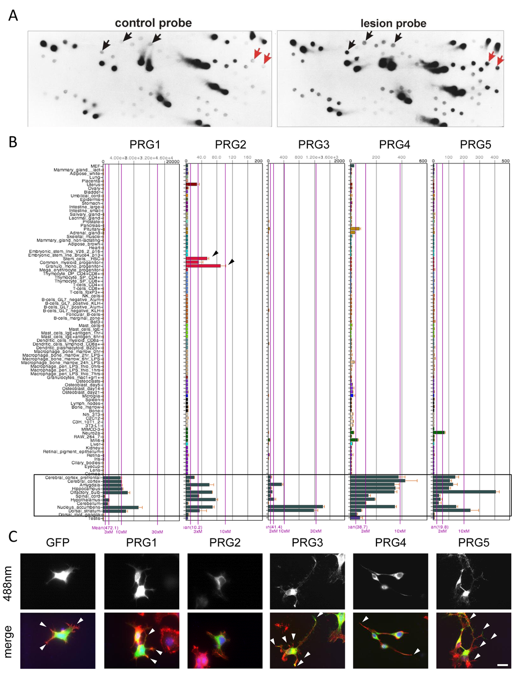
Figure 1.Differential subtractive cDNA library screening reveals Plasticity-Related genes (PRGs) with neuronal cell morphology challenging effects. (A) Representative differential subtractive cDNA dot blot library screening for lesion-induced genes in the hippocampus. Duplicative dot blots were probed with cDNAs from controls (adult hippocampus cDNAs, left) and lesioned brains (differentiated hippocampus cDNAs, right). Note that some genes are solely detectable with probes from lesioned hippocampus (red arrows), whereas others are quantitatively regulated (black arrows). (B) BIO-GPS organ and tissue expression analysis of PRG1-5 (left to right) in the mouse. Note, that expression of PRGs is preferably high in different brain areas (black framed box). PRG1 is expressed highest with an average of 472.1 and PRG2 is lowest with 10.1 in average (average compared to overall mean expression of individual array). Notably, PRG2 is also expressed in myeloid progenitors (black arrowheads). (C) Comparative analysis of GFP and various PRG family members in (GFP and PRG1 to 5-GFP, left to right) in murine neuronal cells N1E-115. Overexpression of PRG3 and PRG5 in particular enhanced filopodia formation and neurite outgrowth (white arrowheads). Expression of PRG1 and PRG2 do not challenge the neuronal phenotype compared to controls. PRG4 induces a bipolar phenotype (arrowheads). Scale bar represents 20 µm.