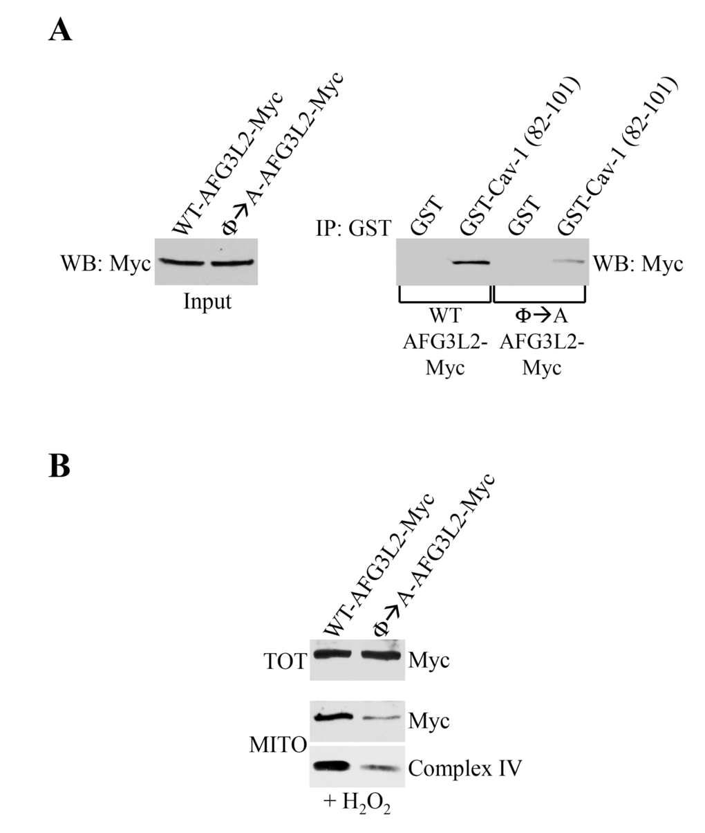
Figure 7.Φ→A-AFG3L2 poorly interacts with caveolin-1, does not accumulate in mitochondria and promotes degradation of complex IV after oxidative stress. (A) GST-Cav-1(82-101) was used in pull down assays with cell lysates from NIH 3T3 cells transiently transfected with either wild type AFG3L2-myc or Φ→A-AFG3L2-myc. Pull-down assays with GST alone was used as internal control. (B) Wild type mouse embryonic fibroblasts (MEFs) were infected with a lentiviral vector (pLVX) expressing either WT-AFG3L2-myc or Φ→A-AFG3L2-myc. After 48 hours, cells were treated with sublethal doses of hydrogen peroxide (150 μM) for 2 hours. Cells were then recovered in complete medium for 7 days. Mitochondrial fractions (MITO) were isolated and the expression levels of AFG3L2-myc and complex IV were measured by immunoblotting analysis. Total expression (TOT) of WT-AFG3L2-myc and Φ→A-AFG3L2-myc is shown in the upper panel.