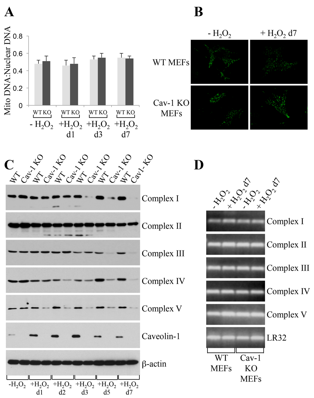
Figure 5.Oxidative stress promotes degradation of mitochondrial respiratory chain complexes in caveolin-1 null MEFs. Wild type and caveolin-1 null mouse embryonic fibroblasts (MEFs) were treated with sublethal doses of hydrogen peroxide (150 μM) for 2 hours. Cells were then recovered in complete medium for different periods of time. Untreated cells (-H2O2) were used as control. (A) The ratio of mitochondrial to nuclear DNA was quantified by performing RT-PCR analysis for the mitochondrial gene ND1 and the nuclear encoded gene Histone 19 using gene-specific primers. (B) Cells were incubated with Mitotracker Green FM (Thermo Fisher Scientific; Waltham, MA) at a concentration of 100 nM in DMEM. Cells were incubated at 37°C for 30 min, washed with PBS and imaged using a Zeiss Confocal Microscope (LSM 5 Pascal; Carl Zeiss, Jena, Germany). (C) The expression level of complex I, complex II, complex III, complex IV, complex V and caveolin-1 was determined by immunoblotting analysis using specific antibody probes. Immunoblotting with anti-β-actin IgGs was performed as internal control. (D) RT-PCR analysis for complex I, complex II, complex III, complex IV and complex V was performed using gene-specific primers. RT-PCR analysis using primers for LR32 was performed as internal control.