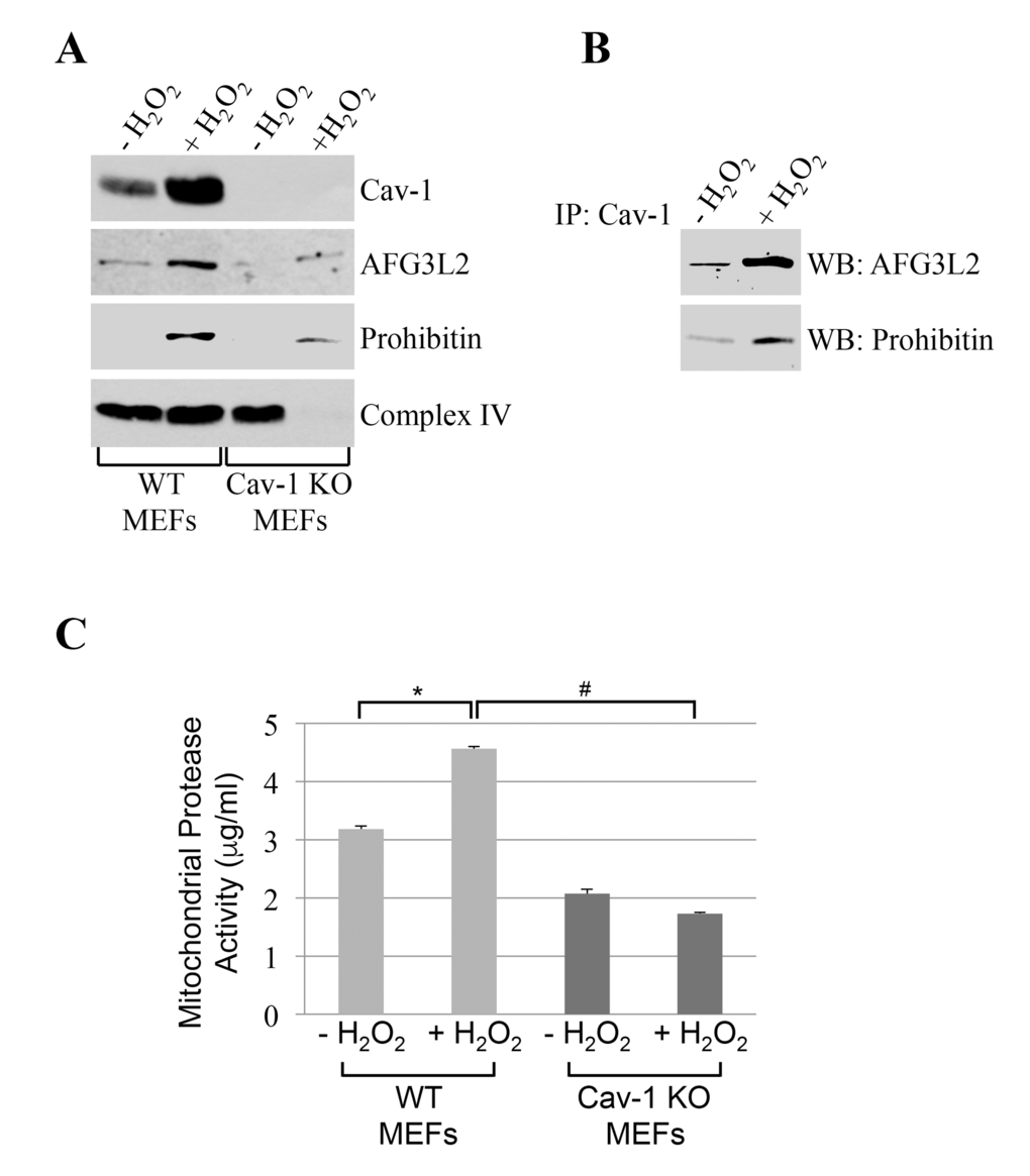
Figure 2.AFG3L2 interacts with caveolin-1 after oxidative stress in vivo. Wild type and caveolin-1 null mouse embryonic fibroblasts (MEFs) were treated with sublethal doses of hydrogen peroxide (150 μM) for 2 hours. Cells were then recovered in complete medium for 7 days. We have chosen these conditions because we have previously shown that they upregulate caveolin-1 expression [20,22,23] and activate caveolin-1-mediated signaling [19,21,24,28,29]. Untreated cells (-H2O2) were used as control. (A) Mitochondrial fractions were isolated and the expression levels of caveolin-1, prohibitin-1 and AFG3L2 were measured by immunoblotting analysis. (B) Mitochondrial fractions were isolated from untreated and hydrogen peroxide-treated wild type MEFs and immunoprecipitated using an antibody probe specific for caveolin-1 (Cav-1); immunoprecipitates were then subjected to immunoblotting analysis with anti-AFG3L2 and prohibitin-1 IgGs. (C) Mitochondria were isolated and mitochondrial protease activity was quantified using the Protease Fluorescent Detection Kit from Sigma-Aldrich (St. Louis, MO) (PF0100). Values in (C) represent mean ± SEM; *,#P<0.001.