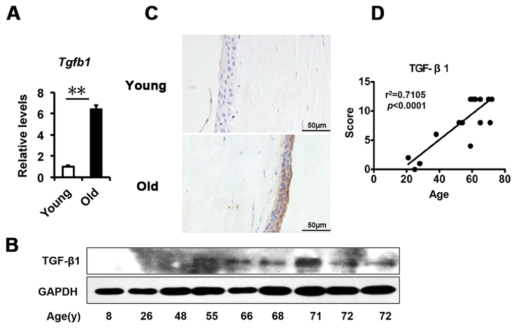
Figure 2.TGF-β1 excess in old donor corneal epithelium. (A) The mRNA expression of Tgfb1 in young donors and old donors corneal epithelium(**P≤0.01, n=3). (B) Immunoblot analysis of TGF-β1 in the corneal epithelium during aging. (C-D) Representative photographs (C) and histopathology scores (D) for the IHC staining of TGF-β1 in corneal epithelium from donors of different ages. There was a statistically significant difference in TGF-β1expression between the donors of younger than 30 years and older than 50years of age (P≤0.01). The figure depicts a Pearson correlation of TGF-β1 expression with age (D).