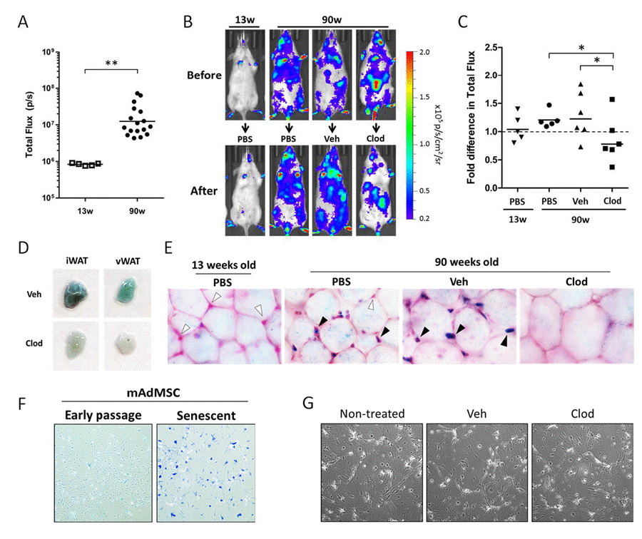
Figure 4.Clodronate treatment depletes p16(Ink4a)-positive and β-galpH6-positive cells from chronologically aged p16LUC mice(A) Bioluminescent baseline readings from the abdomen of young (13 weeks) versus old (90 weeks) p16LUC mice (n=5 and 17 mice/group, respectively). Geometric mean is depicted on graph. (B-C) Old mice were randomized among 3 groups based on bioluminescence from the abdomen (n=5-6 per group): treatment with PBS, vehicle liposomes in PBS (Veh), or liposomal clodronate (Clod). Bioluminescence of the abdomens was measured after two clodronate treatments (i.p., three days prior and i.v., one days prior to luminescent measurement). (B) Representative serial images of p16LUC mice depicting luminescence (in radiance) before and after treatment regimen. Colored scale depicts relative luminescent signal intensity of minimum and maximum thresholds, displayed in terms of radiance. (C) The amount of luminescent signal (total flux; p/s) from the abdomen of treated p16LUC mice is expressed as the fold difference compared to measurement before treatment. Geometric mean is depicted on graph. (D) Inguinal and visceral (perigonadal) depots of white adipose tissue (iWAT and vWAT, respectively) were collected from vehicle and clodronate liposome treated 90-week old p16LUC mice and stained for β-galpH6 activity. Representative photographic images are presented. (E) Representative light microscopy images (magnification, 200x) of β-galpH6-stained visceral adipose tissue counterstained with nuclear fast red. Cells residing between adipocytes (indicated by the presence of nuclear stain) are β-galpH6-negative (white arrow) or -positive (black arrow). These cells are altogether absent from large regions in clodronate-treated mice (as depicted). (F) Representative images of β-galpH6-stained cultures of mouse adipose-derived mesenchymal stromal cells (mAdMSC) from p16LUC mice at early passage (p1 cultures) or 10 days after 20Gy gamma-irradiation (Senescent). SCs stain positive for β-galpH6 and are enlarged and morphologically distinct from early passage. (G) Phase contrast light microscopy images of senescent mAdMSCs following overnight (20 hr) with 50 µg/mL clodronate liposomes (Clod), or similar dilution (1:100) of vehicle liposomes (Veh) or PBS (non-treated), indicating no observable cell death or effects on these cells.