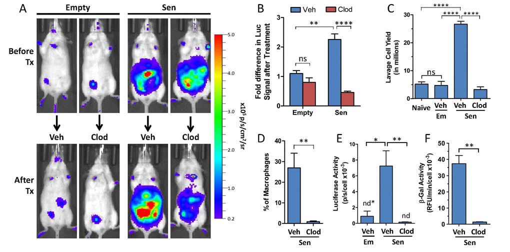
Figure 3.Pharmacological clearance of macrophages in vivo depletes luciferase and β-galpH6 activity from p16LUC mice bearing SCs(A) Representative serial images depicting in vivo bioluminescence from p16LUC mice acquired 12 days after inoculation of empty beads (Empty) or alginate-embedded SCs (before treatment) and one week later (after treatment; 18-20 days post-inoculation) after two i.p. administrations of liposomes containing PBS control (Veh) or clodronate (Clod). Colored scale depicts relative luminescent signal intensity of minimum and maximum thresholds, displayed in terms of radiance. (B) The amount of luminescence (total flux; p/s) from the abdomen after treatment is expressed as the fold difference compared to the signal measured before treatment for each group. (C) Total yield of cells recovered from peritoneal lavage from naïve mice, of liposomal vehicle-treated mice bearing empty beads (Em/Veh), or of liposomal vehicle- or clodronate-treated mice bearing SCs (Sen/Veh and Sen/Clod, respectively). (D) The amount of macrophages present in peritoneal lavage of treated mice bearing SCs is expressed as the percentage of F4/80-positive cells present within the population of live CD45-positive cells, as assessed via flow cytometry on immunostained cells. Cell lysates of whole lavage after treatment were assayed for luciferase activity (E) and β-galpH6 activity (F), normalized to cell number. Values depicted are means +/− SEM (n = 3-7 mice/group). ns = not statistically significant, p>0.05; n.d. = not detectable, values depicted indicate detection limit (defined as 2-fold above background reading) per cell number analyzed.