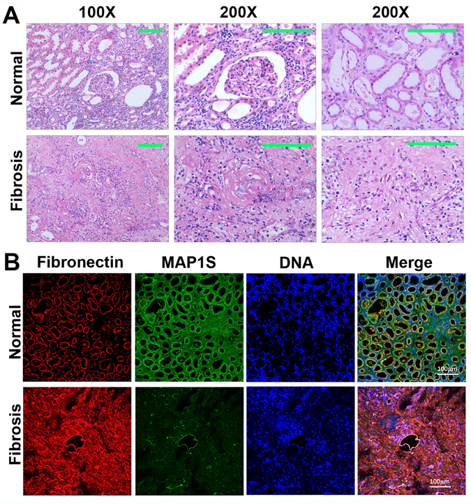
Figure 1.Levels of MAP1S are decreased and levels of fibronectin are elevated in renal tissues from patients suffering from renal fibrosis(A) Representative images showing the H&E staining of renal tissues collected from patients suffering from renal fibrosis and healthy control. The area containing a glomerulus or distal and proximal convoluted tubules is amplified (200X) to show the detail structures. (B) Representative images showing the immuno-florescent staining of MAP1S (green), fibronectin (red) and nuclear DNA (blue) in the normal and renal fibrotic tissues. Scale Bar: 100 μm.