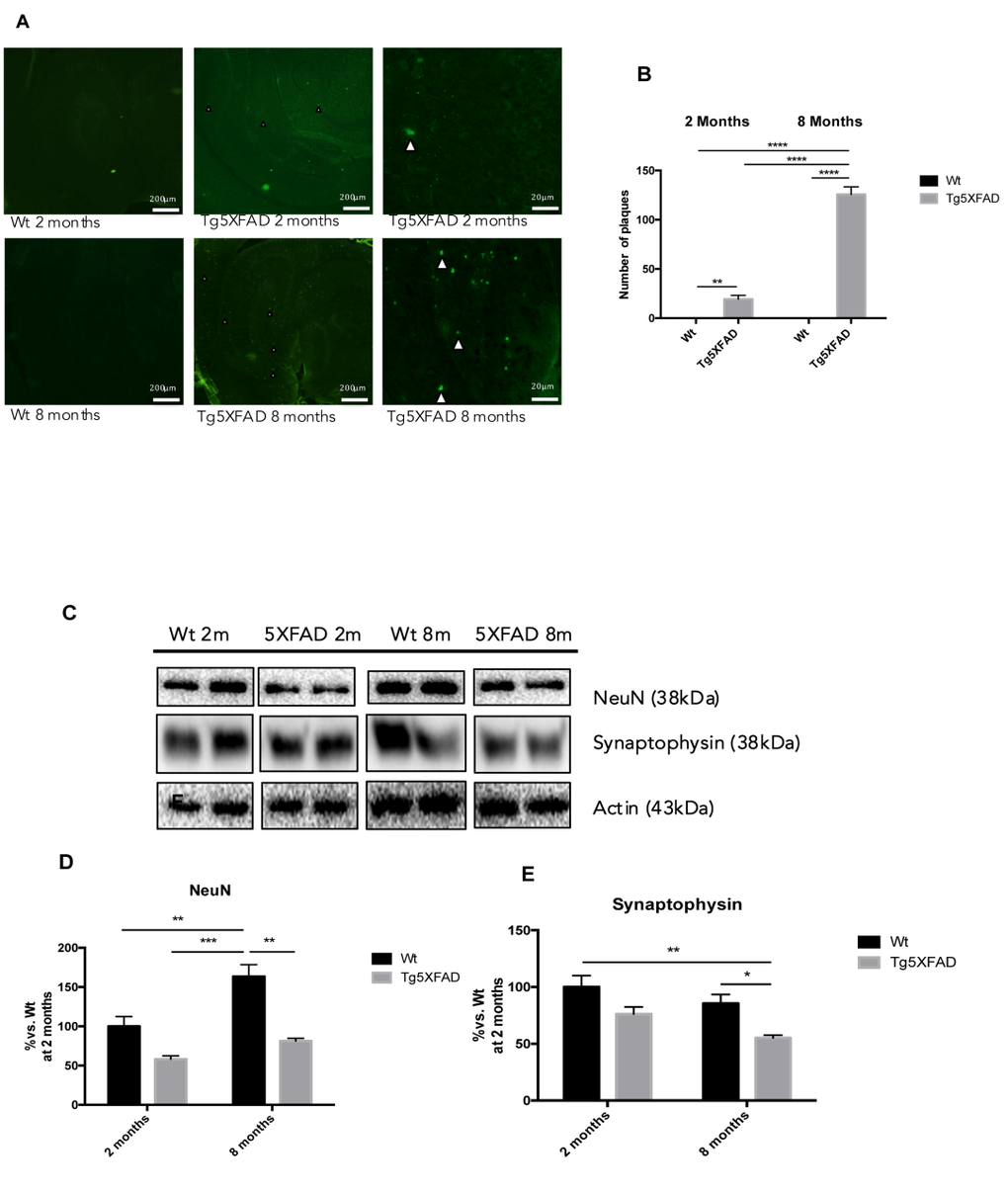
Figure 7.Histological images of amyloid plaques stained with thioflavin-S in female mice aged 2 and 8 months (Wt and 5XFAD). There is a heavy load of plaques in the majority of the brain areas ilustrated in 5XFAD the brain section (A). Representative Western blot for NeuN (B, C) and Synaptophysin (D, E). Bars represent mean ± Standard Error of the Mean (SEM), n = 4 for each group; DG: Dentate Gyrus. Scale bar for histochemical images is indicated in the Picture; *p<0.05; **p<0.01; ***p<0.001.