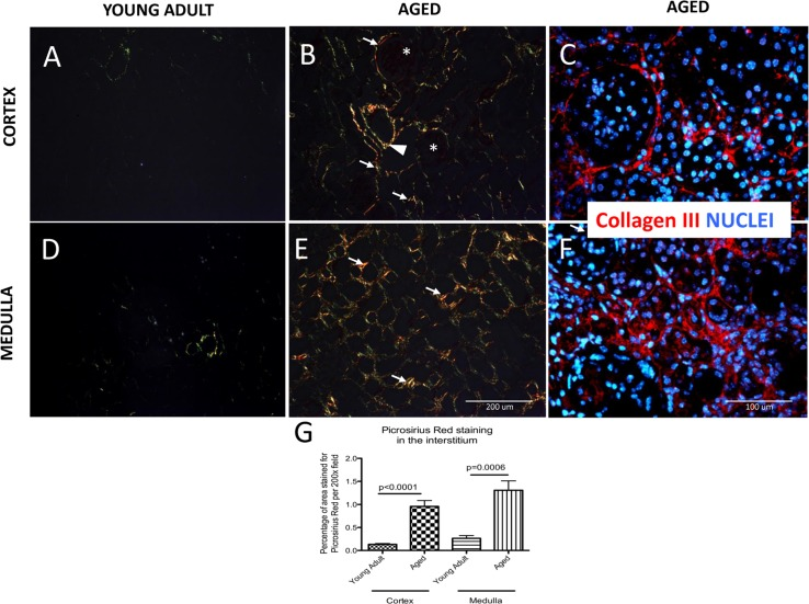
Figure 2.Accumulation of type I and III collagen in aged kidneysPicrosirius Red staining of collagen I and III was examined by polarized light microscopy. Additionally, collagen III fibers are visualized with specific antibody (red color), nuclei are labeled with dapi (blue color). (A) Collagen fibers were sparse in the cortex of young adult kidneys. (B) Aged kidneys demonstrated interstitial collagen deposition (arrows indicate examples) in the peritubular and periglomerular area (glomeruli are indicated by asterix). Adventitia of arterial wall was enriched in collagen (arrowhead). (C) Collagen III staining confirms the location of interstitial and periglomerular fibrosis. (D) Picrosirius Red was barely detected in the medulla of young adult kidneys. (E) Picrosirius Red staining was markedly increased in the medulla of aged mice (arrows indicate examples) in the interstitium. (F) Collagen III staining confirms tubulointerstital fibrosis. (G) Graph of quantitation: Picrosirius Red staining significantly increased in the interstitium in both the cortex and the medulla of aged mice. Data are represented as mean ± SEM (n=6).